Christensen Anders, Juhl Karina, Persson Morten, Charabi Birgitte Wittenborg, Mortensen Jann, Kiss Katalin, Lelkaitis Giedrius, Rubek Niclas, von Buchwald Christian, Kjær Andreas
Department of Otolaryngology, Head & Neck Surgery and Audiology, Rigshospitalet, Copenhagen University Hospital, Denmark.
Department of Clinical Physiology, Nuclear Medicine & PET and Cluster for Molecular Imaging, Rigshospitalet and University of Copenhagen, Denmark.
Oncotarget. 2017 Feb 28;8(9):15407-15419. doi: 10.18632/oncotarget.14282.
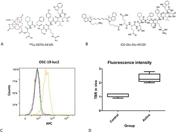
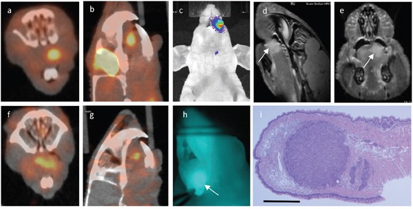
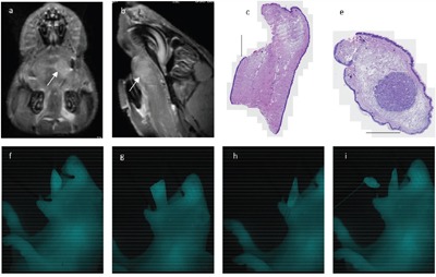
Urokinase-like Plasminogen Activator Receptor (uPAR) is overexpressed in a variety of carcinoma types, and therefore represents an attractive imaging target. The aim of this study was to assess the feasibility of two uPAR-targeted probes for PET and fluorescence tumor imaging in a human xenograft tongue cancer model.
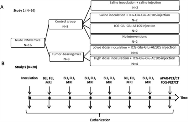
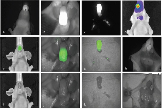
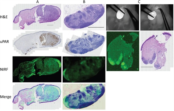
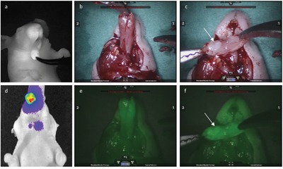
Tumor growth of tongue cancer was monitored by bioluminescence imaging (BLI) and MRI. Either ICG-Glu-Glu-AE105 (fluorescent agent) or 64Cu-DOTA-AE105 (PET agent) was injected systemically, and fluorescence imaging or PET/CT imaging was performed. Tissue was collected for micro-fluorescence imaging and histology. A clear fluorescent signal was detected in the primary tumor with a mean in vivo tumor-to-background ratio of 2.5. Real-time fluorescence-guided tumor resection was possible, and sub-millimeter tumor deposits could be localized. Histological analysis showed co-localization of the fluorescent signal, uPAR expression and tumor deposits. In addition, the feasibility of uPAR-guided robotic cancer surgery was demonstrated. Also, uPAR-PET imaging showed a clear and localized signal in the tongue tumors.
This study demonstrated the feasibility of combining two uPAR-targeted probes in a preclinical head and neck cancer model. The PET modality provided preoperative non-invasive tumor imaging and the optical modality allowed for real-time fluorescence-guided tumor detection and resection. Clinical translation of this platform seems promising.
尿激酶型纤溶酶原激活物受体(uPAR)在多种癌症类型中过表达,因此是一个有吸引力的成像靶点。本研究的目的是评估两种靶向uPAR的探针在人舌癌异种移植模型中用于PET和荧光肿瘤成像的可行性。
通过生物发光成像(BLI)和MRI监测舌癌的肿瘤生长。全身注射ICG-Glu-Glu-AE105(荧光剂)或64Cu-DOTA-AE105(PET剂),然后进行荧光成像或PET/CT成像。收集组织进行微荧光成像和组织学检查。在原发性肿瘤中检测到清晰的荧光信号,体内肿瘤与背景的平均比值为2.5。实时荧光引导下的肿瘤切除是可行的,亚毫米级的肿瘤沉积物可以定位。组织学分析显示荧光信号、uPAR表达和肿瘤沉积物共定位。此外,还证明了uPAR引导的机器人癌症手术的可行性。而且,uPAR-PET成像在舌肿瘤中显示出清晰的局部信号。
本研究证明了在临床前头颈癌模型中联合使用两种靶向uPAR的探针的可行性。PET模式提供术前非侵入性肿瘤成像,光学模式允许实时荧光引导的肿瘤检测和切除。该平台的临床转化似乎很有前景。