• 文献检索
  • 文档翻译
  • 深度研究
  • 学术资讯
  • Suppr Zotero 插件Zotero 插件
  • 邀请有礼
  • 套餐&价格
  • 历史记录
应用&插件
Suppr Zotero 插件Zotero 插件浏览器插件Mac 客户端Windows 客户端微信小程序
定价
高级版会员购买积分包购买API积分包
服务
文献检索文档翻译深度研究API 文档MCP 服务
关于我们
关于 Suppr公司介绍联系我们用户协议隐私条款
关注我们

Suppr 超能文献

核心技术专利:CN118964589B侵权必究
粤ICP备2023148730 号-1Suppr @ 2026

文献检索

告别复杂PubMed语法,用中文像聊天一样搜索,搜遍4000万医学文献。AI智能推荐,让科研检索更轻松。

立即免费搜索

文件翻译

保留排版,准确专业,支持PDF/Word/PPT等文件格式,支持 12+语言互译。

免费翻译文档

深度研究

AI帮你快速写综述,25分钟生成高质量综述,智能提取关键信息,辅助科研写作。

立即免费体验

转化生长因子β激活激酶 1 通过转录抑制乙型肝炎病毒复制。

Transforming growth factor β-activated kinase 1 transcriptionally suppresses hepatitis B virus replication.

机构信息

State Key Laboratory of Organ Failure Research, Guangdong Provincial Key Laboratory of Viral Hepatitis Research, Department of Infectious Diseases, Nanfang Hospital, Southern Medical University, Guangzhou, China.

Institute of Virology, University Hospital of Essen, Essen, Germany.

出版信息

Sci Rep. 2017 Jan 3;7:39901. doi: 10.1038/srep39901.

DOI:10.1038/srep39901
PMID:28045080
原文链接:https://pmc.ncbi.nlm.nih.gov/articles/PMC5206675/
Abstract

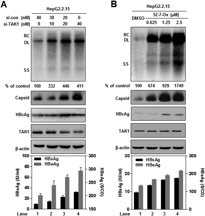
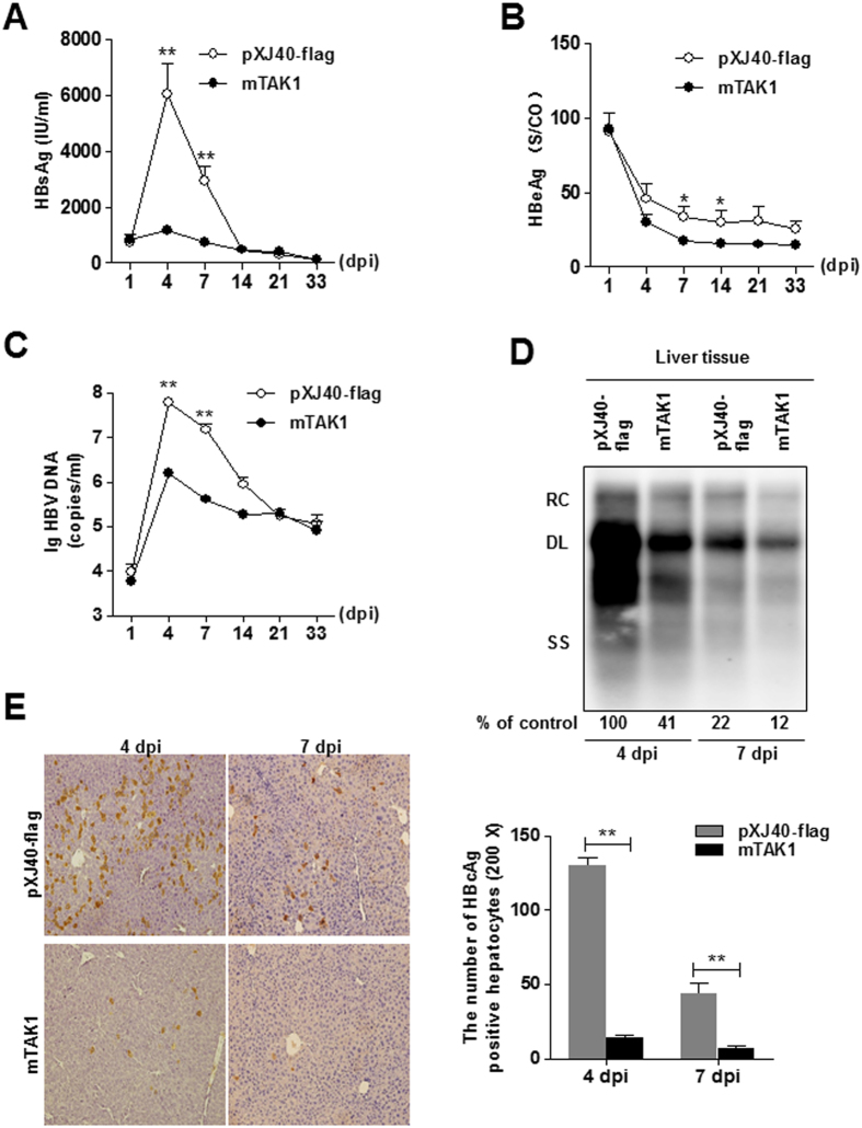
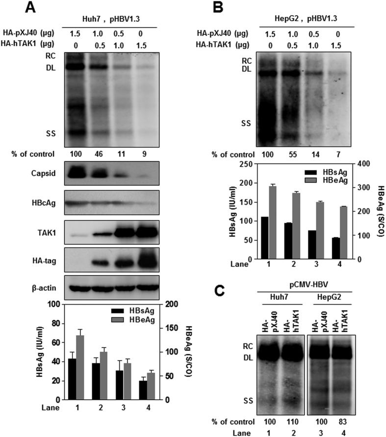
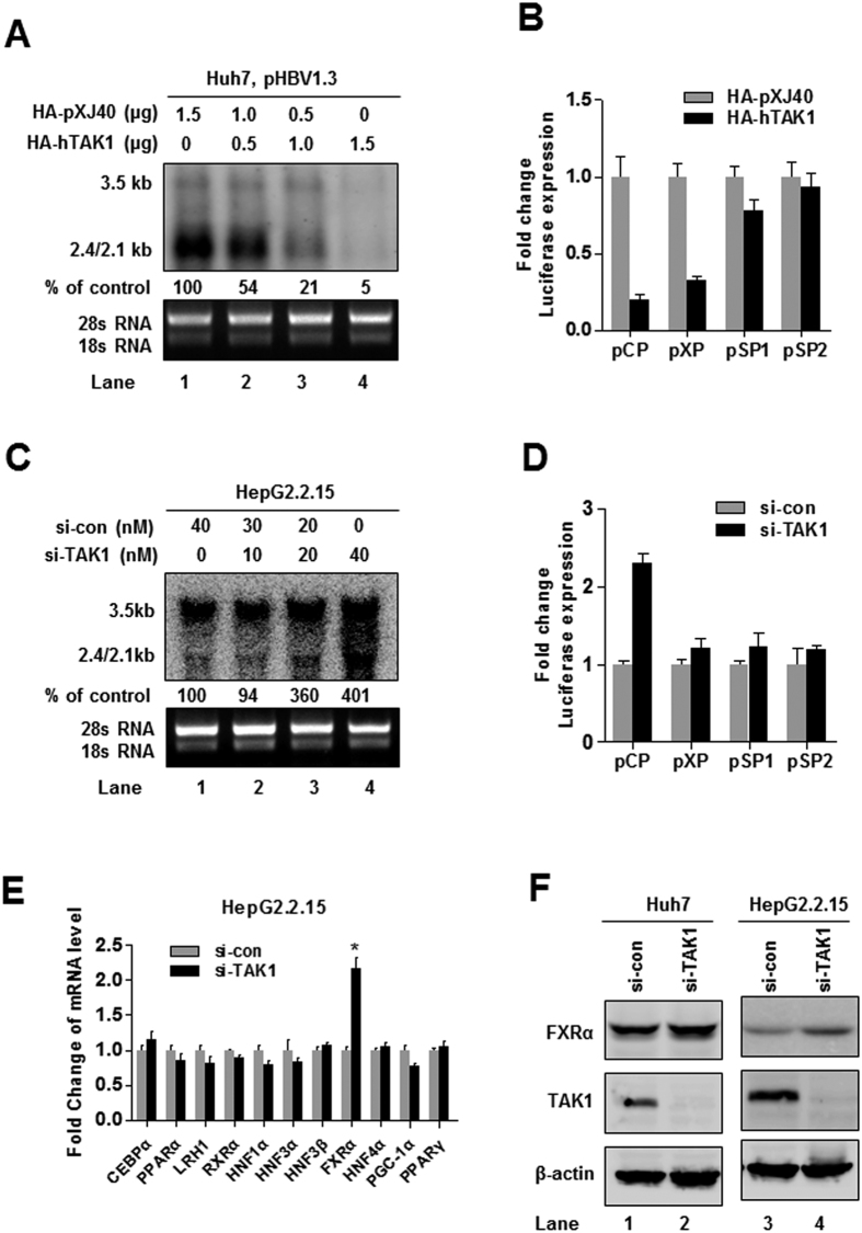
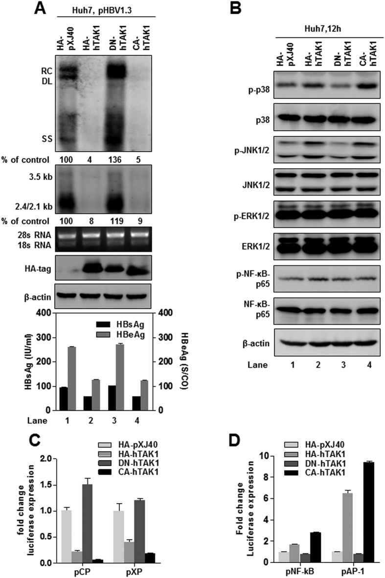
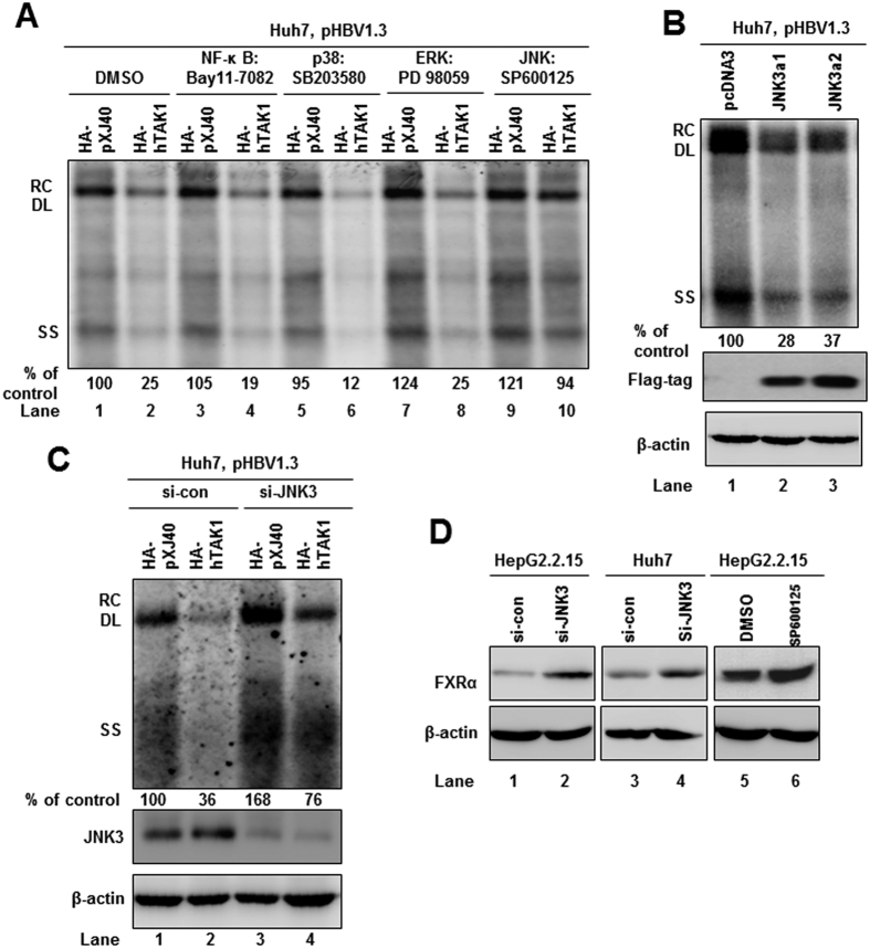
Hepatitis B Virus (HBV) replication in hepatocytes is restricted by the host innate immune system and related intracellular signaling pathways. Transforming growth factor β-activated kinase 1 (TAK1) is a key mediator of toll-like receptors and pro-inflammatory cytokine signaling pathways. Here, we report that silencing or inhibition of endogenous TAK1 in hepatoma cell lines leads to an upregulation of HBV replication, transcription, and antigen expression. In contrast, overexpression of TAK1 significantly suppresses HBV replication, while an enzymatically inactive form of TAK1 exerts no effect. By screening TAK1-associated signaling pathways with inhibitors and siRNAs, we found that the MAPK-JNK pathway was involved in TAK1-mediated HBV suppression. Moreover, TAK1 knockdown or JNK pathway inhibition induced the expression of farnesoid X receptor α, a transcription factor that upregulates HBV transcription. Finally, ectopic expression of TAK1 in a HBV hydrodynamic injection mouse model resulted in lower levels of HBV DNA and antigens in both liver and serum. In conclusion, our data suggest that TAK1 inhibits HBV primarily at viral transcription level through activation of MAPK-JNK pathway, thus TAK1 represents an intrinsic host restriction factor for HBV replication in hepatocytes.

摘要

乙型肝炎病毒 (HBV) 在肝细胞中的复制受到宿主固有免疫系统和相关细胞内信号通路的限制。转化生长因子β激活激酶 1 (TAK1) 是 Toll 样受体和促炎细胞因子信号通路的关键介质。在这里,我们报告称,在肝癌细胞系中沉默或抑制内源性 TAK1 会导致 HBV 复制、转录和抗原表达上调。相比之下,TAK1 的过表达可显著抑制 HBV 复制,而具有酶活性的 TAK1 则没有影响。通过用抑制剂和 siRNA 筛选与 TAK1 相关的信号通路,我们发现 MAPK-JNK 通路参与了 TAK1 介导的 HBV 抑制。此外,TAK1 敲低或 JNK 通路抑制诱导法尼酯 X 受体 α 的表达,法尼酯 X 受体 α 是一种上调 HBV 转录的转录因子。最后,在 HBV 动力学注射小鼠模型中异位表达 TAK1 会导致肝和血清中 HBV DNA 和抗原水平降低。总之,我们的数据表明,TAK1 通过激活 MAPK-JNK 通路主要在病毒转录水平上抑制 HBV,因此 TAK1 代表了肝细胞中 HBV 复制的固有宿主限制因子。

https://cdn.ncbi.nlm.nih.gov/pmc/blobs/761c/5206675/abf1d1bdc6a0/srep39901-f6.jpg
https://cdn.ncbi.nlm.nih.gov/pmc/blobs/761c/5206675/5c21e4fbf97b/srep39901-f1.jpg
https://cdn.ncbi.nlm.nih.gov/pmc/blobs/761c/5206675/41dbc676024a/srep39901-f2.jpg
https://cdn.ncbi.nlm.nih.gov/pmc/blobs/761c/5206675/d4d47d468e48/srep39901-f3.jpg
https://cdn.ncbi.nlm.nih.gov/pmc/blobs/761c/5206675/7f9d4f99fc00/srep39901-f4.jpg
https://cdn.ncbi.nlm.nih.gov/pmc/blobs/761c/5206675/e05d93fde3c9/srep39901-f5.jpg
https://cdn.ncbi.nlm.nih.gov/pmc/blobs/761c/5206675/abf1d1bdc6a0/srep39901-f6.jpg

相似文献

1
Transforming growth factor β-activated kinase 1 transcriptionally suppresses hepatitis B virus replication.转化生长因子β激活激酶 1 通过转录抑制乙型肝炎病毒复制。
Sci Rep. 2017 Jan 3;7:39901. doi: 10.1038/srep39901.
2
Stimulation of TRAF6/TAK1 degradation and inhibition of JNK/AP-1 signalling by ginsenoside Rg3 attenuates hepatitis B virus replication.人参皂苷 Rg3 通过降解 TRAF6/TAK1 和抑制 JNK/AP-1 信号通路来抑制乙型肝炎病毒复制。
Int J Biochem Cell Biol. 2013 Nov;45(11):2612-21. doi: 10.1016/j.biocel.2013.08.016. Epub 2013 Sep 1.
3
Farnesoid X receptor-α is a proviral host factor for hepatitis B virus that is inhibited by ligands in vitro and in vivo.法尼醇 X 受体-α是乙型肝炎病毒的前病毒宿主因子,在体外和体内可被配体抑制。
FASEB J. 2019 Feb;33(2):2472-2483. doi: 10.1096/fj.201801181R. Epub 2018 Oct 11.
4
Role of Toll-like receptor 2 in the immune response against hepadnaviral infection.Toll 样受体 2 在抗病毒感染免疫反应中的作用。
J Hepatol. 2012 Sep;57(3):522-8. doi: 10.1016/j.jhep.2012.05.004. Epub 2012 May 19.
5
Hepatitis B virus suppresses toll-like receptor-mediated innate immune responses in murine parenchymal and nonparenchymal liver cells.乙肝病毒抑制小鼠肝实质细胞和非实质细胞中 toll 样受体介导的天然免疫反应。
Hepatology. 2009 Apr;49(4):1132-40. doi: 10.1002/hep.22751.
6
Hepatitis B Virus Stimulated Fibronectin Facilitates Viral Maintenance and Replication through Two Distinct Mechanisms.乙肝病毒刺激的纤连蛋白通过两种不同机制促进病毒维持和复制。
PLoS One. 2016 Mar 29;11(3):e0152721. doi: 10.1371/journal.pone.0152721. eCollection 2016.
7
Mucroporin-M1 inhibits hepatitis B virus replication by activating the mitogen-activated protein kinase (MAPK) pathway and down-regulating HNF4α in vitro and in vivo.粘蛋白 M1 通过激活丝裂原活化蛋白激酶(MAPK)通路和下调体内外 HNF4α 抑制乙型肝炎病毒复制。
J Biol Chem. 2012 Aug 31;287(36):30181-90. doi: 10.1074/jbc.M112.370312. Epub 2012 Jul 12.
8
Hepatitis B Surface Antigen Suppresses the Activation of Nuclear Factor Kappa B Pathway Interaction With the TAK1-TAB2 Complex.乙型肝炎表面抗原抑制核因子-κB 通路的激活与 TAK1-TAB2 复合物的相互作用。
Front Immunol. 2021 Feb 25;12:618196. doi: 10.3389/fimmu.2021.618196. eCollection 2021.
9
The Functional and Antiviral Activity of Interferon Alpha-Inducible IFI6 Against Hepatitis B Virus Replication and Gene Expression.干扰素诱导的 IFI6 对乙型肝炎病毒复制和基因表达的功能和抗病毒活性。
Front Immunol. 2021 Apr 1;12:634937. doi: 10.3389/fimmu.2021.634937. eCollection 2021.
10
Hepatitis B virus hijacks CTHRC1 to evade host immunity and maintain replication.乙型肝炎病毒劫持 CTHRC1 以逃避宿主免疫并维持复制。
J Mol Cell Biol. 2015 Dec;7(6):543-56. doi: 10.1093/jmcb/mjv048. Epub 2015 Jul 15.

引用本文的文献

1
Hepatitis B Surface Antigen Suppresses the Activation of Nuclear Factor Kappa B Pathway Interaction With the TAK1-TAB2 Complex.乙型肝炎表面抗原抑制核因子-κB 通路的激活与 TAK1-TAB2 复合物的相互作用。
Front Immunol. 2021 Feb 25;12:618196. doi: 10.3389/fimmu.2021.618196. eCollection 2021.
2
ATP1B3 cooperates with BST-2 to promote hepatitis B virus restriction.ATP1B3 与 BST-2 合作促进乙型肝炎病毒限制。
J Med Virol. 2020 Feb;92(2):201-209. doi: 10.1002/jmv.25599. Epub 2019 Oct 16.
3
Pre-Activation of Toll-Like Receptor 2 Enhances CD8 T-Cell Responses and Accelerates Hepatitis B Virus Clearance in the Mouse Models.

本文引用的文献

1
Antiviral activity of various interferons and pro-inflammatory cytokines in non-transformed cultured hepatocytes infected with hepatitis B virus.多种干扰素和促炎细胞因子对感染乙型肝炎病毒的未转化培养肝细胞的抗病毒活性。
Antiviral Res. 2016 Jun;130:36-45. doi: 10.1016/j.antiviral.2016.03.008. Epub 2016 Mar 10.
2
Global strategies are required to cure and eliminate HBV infection.需要采取全球策略来治愈和消除乙肝病毒感染。
Nat Rev Gastroenterol Hepatol. 2016 Apr;13(4):239-48. doi: 10.1038/nrgastro.2016.7. Epub 2016 Feb 24.
3
Interferon-γ and Tumor Necrosis Factor-α Produced by T Cells Reduce the HBV Persistence Form, cccDNA, Without Cytolysis.
Toll样受体2的预激活增强小鼠模型中CD8 T细胞反应并加速乙型肝炎病毒清除
Front Immunol. 2018 Jun 29;9:1495. doi: 10.3389/fimmu.2018.01495. eCollection 2018.
4
Mitogen-activated protein kinase eight polymorphisms are associated with immune responsiveness to HBV vaccinations in infants of HBsAg(+)/HBeAg(-) mothers.丝裂原活化蛋白激酶 8 多态性与 HBsAg(+)/HBeAg(-)母亲的婴儿对乙肝疫苗免疫反应有关。
BMC Infect Dis. 2018 Jun 14;18(1):274. doi: 10.1186/s12879-018-3166-x.
5
Interaction between Hepatitis B Virus and Toll-Like Receptors: Current Status and Potential Therapeutic Use for Chronic Hepatitis B.乙型肝炎病毒与Toll样受体之间的相互作用:慢性乙型肝炎的现状及潜在治疗用途
Vaccines (Basel). 2018 Jan 16;6(1):6. doi: 10.3390/vaccines6010006.
6
Interleukin-35 Level Is Elevated in Patients with Chronic Hepatitis B Virus Infection.白细胞介素-35 水平在慢性乙型肝炎病毒感染患者中升高。
Int J Med Sci. 2018 Jan 1;15(2):188-194. doi: 10.7150/ijms.21957. eCollection 2018.
7
Fucoidan from Fucus vesiculosus suppresses hepatitis B virus replication by enhancing extracellular signal-regulated Kinase activation.岩藻聚糖硫酸酯(褐藻糖胶)通过增强细胞外信号调节激酶的激活来抑制乙肝病毒复制。
Virol J. 2017 Sep 16;14(1):178. doi: 10.1186/s12985-017-0848-8.
T 细胞产生的干扰素-γ 和肿瘤坏死因子-α 可减少 HBV 持续存在形式 cccDNA,而不引起细胞溶解。
Gastroenterology. 2016 Jan;150(1):194-205. doi: 10.1053/j.gastro.2015.09.026. Epub 2015 Sep 28.
4
Estimations of worldwide prevalence of chronic hepatitis B virus infection: a systematic review of data published between 1965 and 2013.全球慢性乙型肝炎病毒感染患病率的估计:1965 年至 2013 年发表数据的系统评价。
Lancet. 2015 Oct 17;386(10003):1546-55. doi: 10.1016/S0140-6736(15)61412-X. Epub 2015 Jul 28.
5
Host-virus interactions in hepatitis B virus infection.乙型肝炎病毒感染中的宿主-病毒相互作用。
Curr Opin Immunol. 2015 Oct;36:61-6. doi: 10.1016/j.coi.2015.06.016. Epub 2015 Jul 15.
6
Molecular biology of hepatitis B virus infection.乙型肝炎病毒感染的分子生物学
Virology. 2015 May;479-480:672-86. doi: 10.1016/j.virol.2015.02.031. Epub 2015 Mar 7.
7
Farnesoid X receptor antagonizes JNK signaling pathway in liver carcinogenesis by activating SOD3.法尼酯X受体通过激活超氧化物歧化酶3(SOD3)拮抗肝脏致癌过程中的JNK信号通路。
Mol Endocrinol. 2015 Feb;29(2):322-31. doi: 10.1210/me.2014-1225. Epub 2014 Dec 12.
8
Cell type-specific function of TAK1 in innate immune signaling.TAK1 在先天免疫信号中的细胞类型特异性功能。
Trends Immunol. 2013 Jul;34(7):307-16. doi: 10.1016/j.it.2013.03.007. Epub 2013 May 7.
9
Transforming growth factor-β signaling in hepatocytes promotes hepatic fibrosis and carcinogenesis in mice with hepatocyte-specific deletion of TAK1.肝细胞中转化生长因子-β信号的转导促进了 TAK1 肝细胞特异性缺失小鼠的肝纤维化和肝癌发生。
Gastroenterology. 2013 May;144(5):1042-1054.e4. doi: 10.1053/j.gastro.2013.01.056. Epub 2013 Feb 4.
10
Sodium taurocholate cotransporting polypeptide is a functional receptor for human hepatitis B and D virus.牛磺胆酸钠共转运多肽是乙型肝炎病毒和丁型肝炎病毒的功能性受体。
Elife. 2012 Nov 13;1:e00049. doi: 10.7554/eLife.00049.