Bu Lingguo, Zhang Ming, Li Jianfeng, Li Fangyi, Liu Heshan, Li Zengyong
Key Laboratory of High Efficiency and Clean Mechanical Manufacture, School of Mechanical Engineering, Shandong University, Jinan, P.R. China.
Interdisciplinary Division of Biomedical Engineering, Faculty of Engineering, The Hong Kong Polytechnic University, Kowloon, Hong Kong, SAR P.R. China.
PLoS One. 2017 Jan 3;12(1):e0169279. doi: 10.1371/journal.pone.0169279. eCollection 2017.
To reveal the physiological mechanism of the decline in cognitive function after sleep deprivation, a within-subject study was performed to assess sleep deprivation effects on phase synchronization, as revealed by wavelet phase coherence (WPCO) analysis of prefrontal tissue oxyhemoglobin signals.
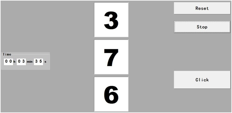
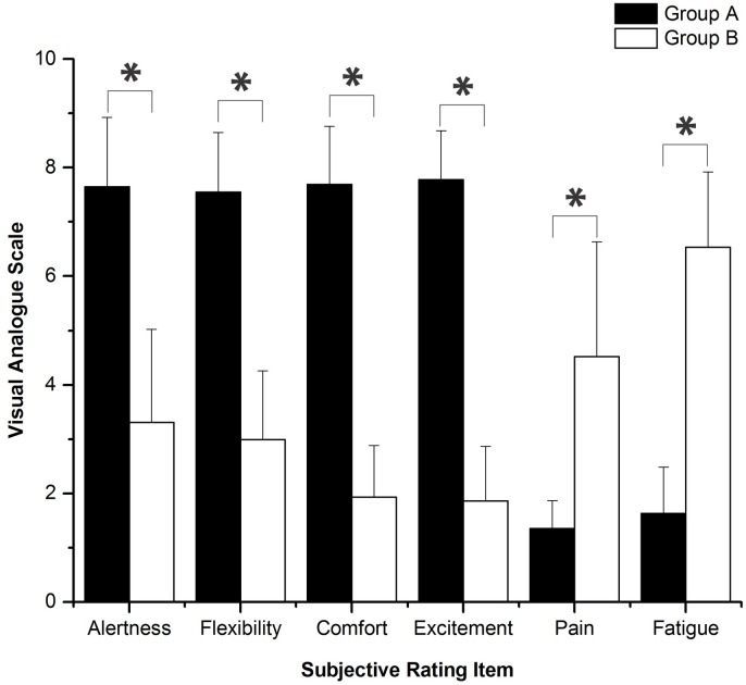
Twenty subjects (10 male and 10 female, 25.5 ± 3.5 years old) were recruited to participate in two tests: one without sleep deprivation (group A) and the other with 24 h of sleep deprivation (group B). Before the test, each subject underwent a subjective evaluation using visual analog scales. A cognitive task was performed by judging three random numbers. Continuous recordings of the near-infrared spectroscopy (NIRS) signals were obtained from both the left and right prefrontal lobes during rest, task, and post-task periods. The WPCO of cerebral Delta [HbO2] signals were analyzed for these three periods for both groups A and B.
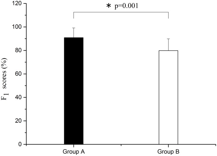
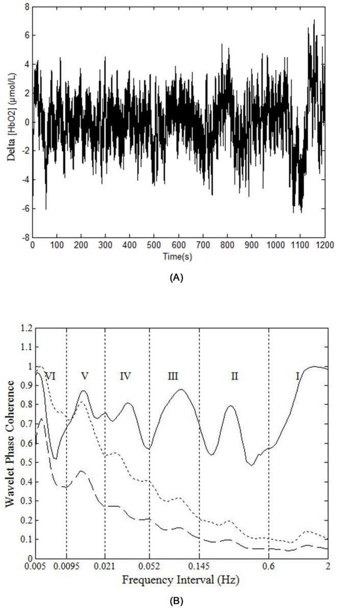
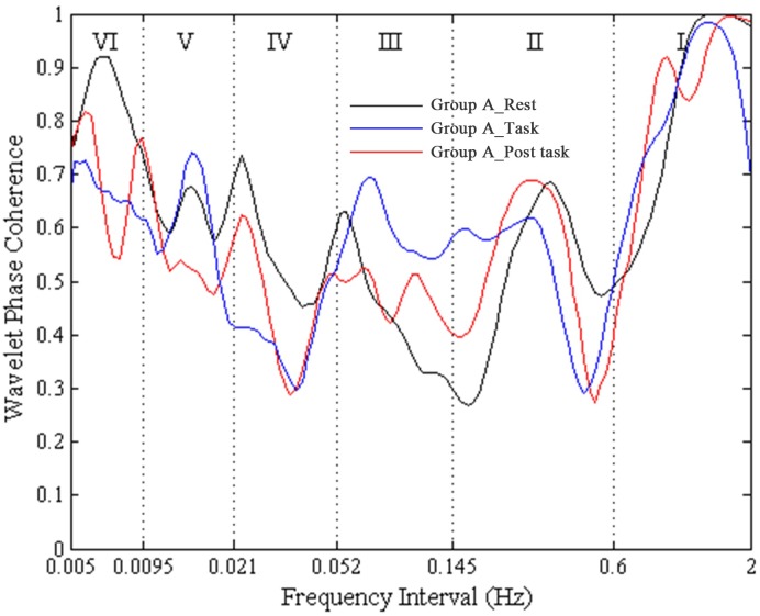
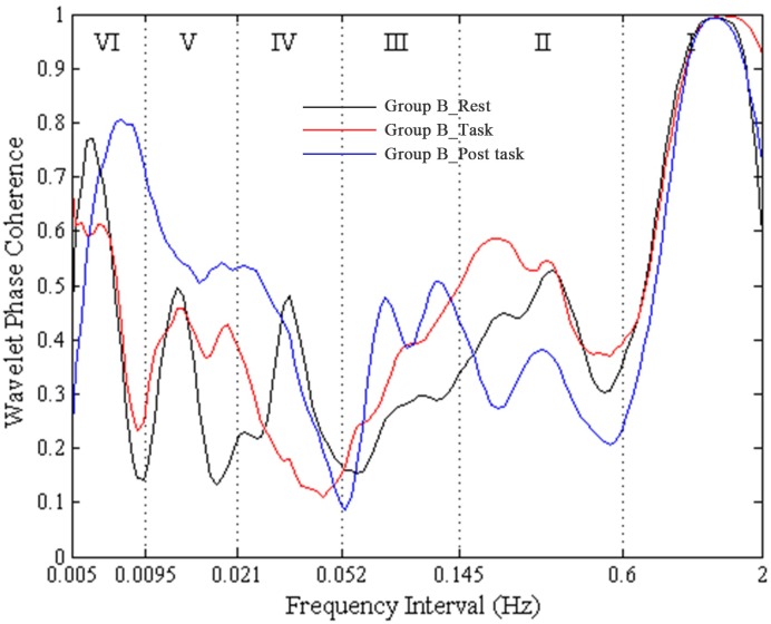
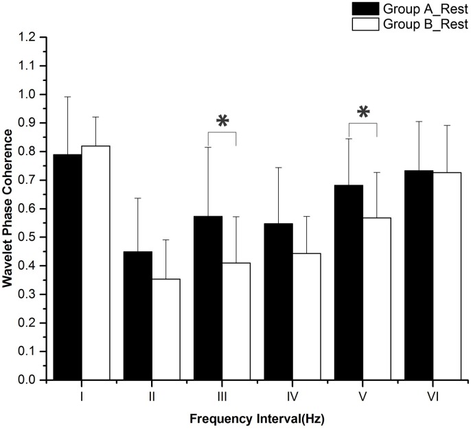
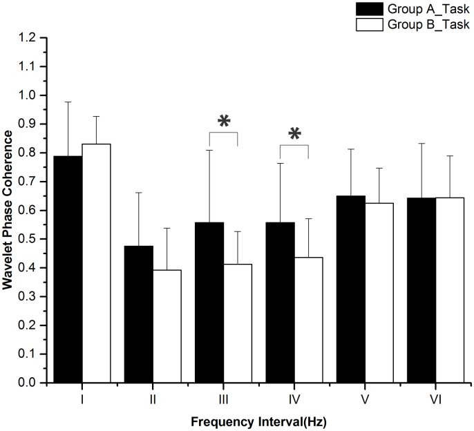
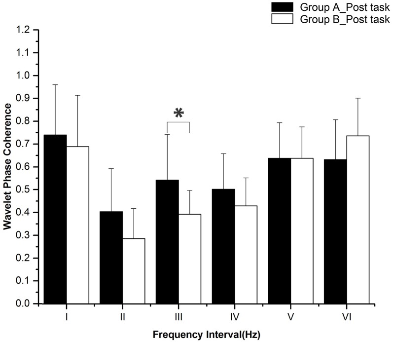
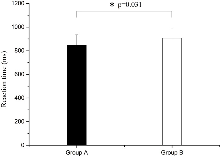
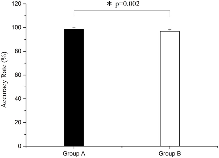
Six frequency intervals were defined: I: 0.6-2 Hz (cardiac activity), II: 0.145-0.6 Hz (respiratory activity), III: 0.052-0.145 Hz (myogenic activity), IV: 0.021-0.052 Hz (neurogenic activity), V: 0.0095-0.021 Hz (nitric oxide related endothelial activity) and VI: 0.005-0.0095 Hz (non-nitric oxide related endothelial activity). WPCO in intervals III (F = 5.955, p = 0.02) and V (F = 4.7, p = 0.037) was significantly lower in group B than in group A at rest. During the task period, WPCO in intervals III (F = 5.175, p = 0.029) and IV (F = 4.585, p = 0.039) was significantly lower in group B compared with group A. In the post-task recovery period, the WPCO in interval III (F = 6.125, p = 0.02) was significantly lower in group B compared with group A. Reaction time was significantly prolonged, and the accuracy rate and F1 score both declined after sleep deprivation.
The decline in WPCO after sleep deprivation indicates reduced phase synchronization between left and right prefrontal oxyhemoglobin oscillations, which may contribute to the diminished cognitive function.
为揭示睡眠剥夺后认知功能下降的生理机制,进行了一项受试者内研究,以评估睡眠剥夺对相位同步的影响,这通过对前额叶组织氧合血红蛋白信号进行小波相位相干(WPCO)分析来揭示。
招募20名受试者(10名男性和10名女性,年龄25.5±3.5岁)参与两项测试:一项无睡眠剥夺(A组),另一项有24小时睡眠剥夺(B组)。测试前,每个受试者使用视觉模拟量表进行主观评估。通过判断三个随机数来执行认知任务。在休息、任务和任务后阶段,从左右前额叶连续记录近红外光谱(NIRS)信号。对A组和B组这三个阶段的脑Delta[HbO2]信号的WPCO进行分析。
定义了六个频率区间:I:0.6 - 2Hz(心脏活动),II:0.145 - 0.6Hz(呼吸活动),III:0.052 - 0.145Hz(肌源性活动),IV:0.021 - 0.052Hz(神经源性活动),V:0.0095 - 0.021Hz(一氧化氮相关内皮活动)和VI:0.005 - 0.0095Hz(非一氧化氮相关内皮活动)。在休息时,B组III区间(F = 5.955,p = 0.02)和V区间(F = 4.7,p = 0.037)的WPCO显著低于A组。在任务期间,B组III区间(F = 5.175,p = 0.029)和IV区间(F = 4.585,p = 0.039)的WPCO显著低于A组。在任务后恢复期,B组III区间(F = 6.125,p = 0.02)的WPCO显著低于A组。睡眠剥夺后反应时间显著延长,准确率和F1分数均下降。
睡眠剥夺后WPCO的下降表明左右前额叶氧合血红蛋白振荡之间的相位同步减少,这可能导致认知功能下降。