Larabee Chelsea M, Desai Shruti, Agasing Agnieshka, Georgescu Constantin, Wren Jonathan D, Axtell Robert C, Plafker Scott M
Oklahoma Center for Neuroscience, University of Oklahoma Health Sciences Center, Oklahoma City, OK; Aging and Metabolism Research Program, Oklahoma Medical Research Foundation, Oklahoma City, OK.
Aging and Metabolism Research Program, Oklahoma Medical Research Foundation, Oklahoma City, OK.
Mol Vis. 2016 Dec 30;22:1503-1513. eCollection 2016.
Optic neuritis, inflammation of the optic nerve, is experienced by most patients with multiple sclerosis (MS) and is typically characterized by episodes of acute, monocular vision loss. These episodes of inflammation can lead to damage or degeneration of the retinal ganglion cells (RGCs), the axons of which comprise the optic nerve. Experimental autoimmune encephalomyelitis (EAE) is a well-established model of MS in which mice are immunized to produce a neuroautoimmunity that recapitulates the cardinal hallmarks of human disease, namely, inflammation, demyelination, and neurodegeneration of the brain, spinal cord, and optic nerve. Inflammation-associated oxidative stress plays a key role in promoting spinal cord damage in EAE. However, the role of oxidative stress in optic neuritis and the associated visual deficits has not been studied. To address this gap in research, we sought to determine how a deficiency in the master antioxidant transcription factor (using nuclear factor-E2-related factor [Nrf2]-deficient mice) affects visual pathology in the EAE model.
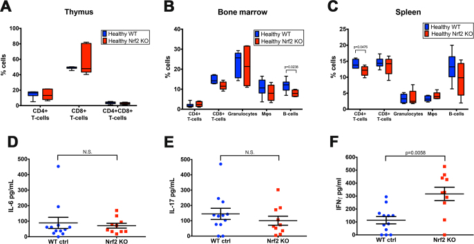
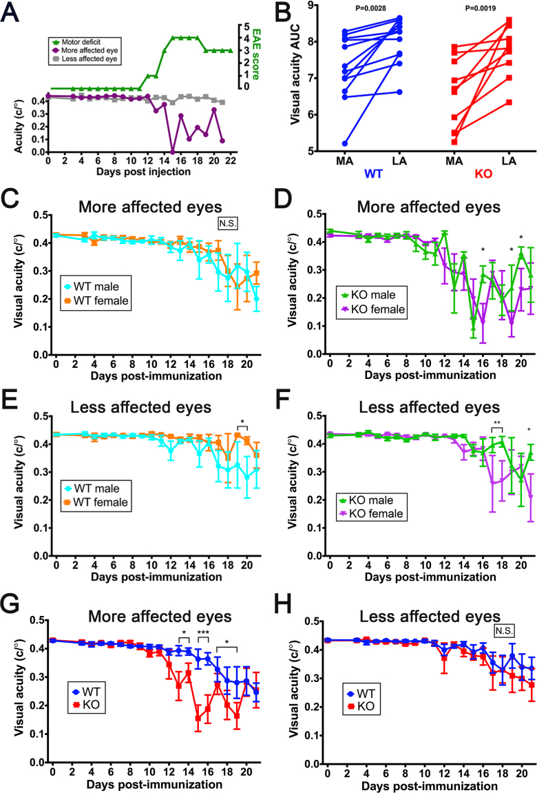
EAE was induced in 8-week-old wild-type (WT) and Nrf2 knockout (KO) mice by immunization against the myelin oligodendrocyte glycoprotein (MOG) peptide antigen. Motor deficits were monitored daily, as was visual acuity using the established functional optokinetic tracking (OKT) assay. Mice were euthanized 21 days post-immunization for histological analyses. The optic nerves were paraffin-embedded and stained with hematoxylin and eosin (H&E) or immune cell type-specific antibodies to analyze inflammatory infiltrates. The retinas were flatmounted and stained with an RGC-specific antibody, and the RGCs were counted to assess neurodegeneration. T-helper (Th) cell-associated cytokines were measured in spleens with enzyme-linked immunosorbent assay (ELISA). Immune analyses of healthy, non-EAE mice were characterized with flow cytometry to assess the baseline immune cell profiles.
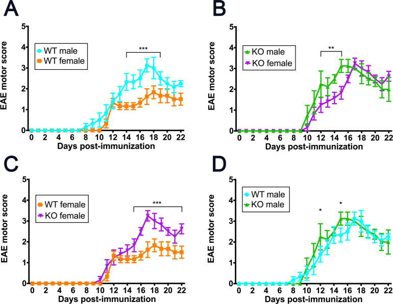
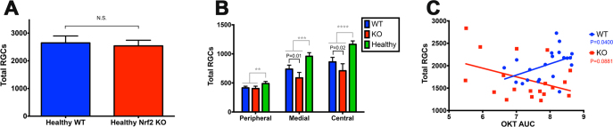
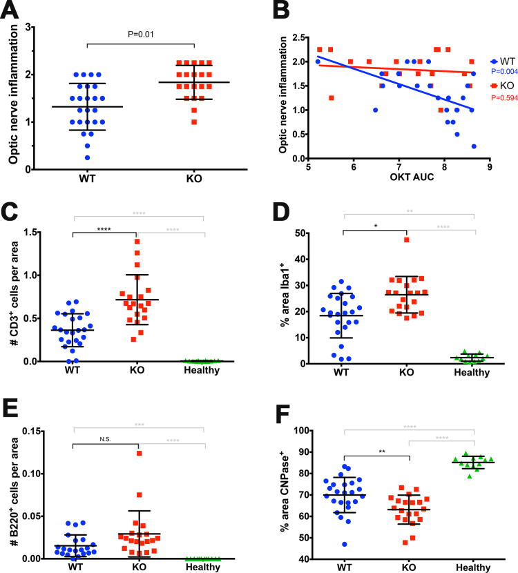
Female Nrf2 KO mice exhibited more severe EAE-induced motor deficits compared with female WT mice. In both genders, EAE elicited more severe visual acuity deficits, inflammation of the optic nerve, and RGC degeneration in KO mice compared with their strain- and age-matched WT counterparts. Visual acuity deficits were primarily present in (and only exacerbated in) one eye of each mouse. Excess inflammatory cells within the optic nerves of the KO mice were primarily comprised of T-cells, and greater RGC degeneration in the KO mice was most prevalent in the central retina compared with the peripheral retina. Nrf2 KO spleens exhibited an increased Th1- but not Th17-associated immune response. This enhanced pathology in the KO mice was not due to global differences in immune system development between the two genotypes.
This is the first study to report that genetic ablation of Nrf2 exacerbates visual deficits, inflammation of the optic nerve, and RGC degeneration in a murine model of MS, suggesting that Nrf2 plays a neuro- and cytoprotective role in EAE-associated optic neuritis.
视神经炎是指视神经的炎症,大多数多发性硬化症(MS)患者都会经历,其典型特征是急性单眼视力丧失发作。这些炎症发作可导致视网膜神经节细胞(RGCs)受损或退化,其轴突构成视神经。实验性自身免疫性脑脊髓炎(EAE)是一种成熟的MS模型,在该模型中,小鼠被免疫以产生一种神经自身免疫,重现人类疾病的主要特征,即大脑、脊髓和视神经的炎症、脱髓鞘和神经变性。炎症相关的氧化应激在促进EAE脊髓损伤中起关键作用。然而,氧化应激在视神经炎及相关视觉缺陷中的作用尚未得到研究。为了填补这一研究空白,我们试图确定主要抗氧化转录因子缺乏(使用核因子-E2相关因子[Nrf2]缺陷小鼠)如何影响EAE模型中的视觉病理。
通过针对髓鞘少突胶质细胞糖蛋白(MOG)肽抗原进行免疫,在8周龄的野生型(WT)和Nrf2基因敲除(KO)小鼠中诱导EAE。每天监测运动功能缺陷,并使用既定的功能性视动跟踪(OKT)试验监测视力。免疫后21天对小鼠实施安乐死以进行组织学分析。将视神经石蜡包埋,并用苏木精和伊红(H&E)或免疫细胞类型特异性抗体染色,以分析炎症浸润。将视网膜铺片并用RGC特异性抗体染色,对RGC进行计数以评估神经变性。用酶联免疫吸附测定(ELISA)测量脾脏中辅助性T(Th)细胞相关细胞因子。对健康的非EAE小鼠进行免疫分析,用流式细胞术评估基线免疫细胞谱。
与雌性WT小鼠相比,雌性Nrf2 KO小鼠表现出更严重的EAE诱导的运动功能缺陷。在两种性别中,与同品系、年龄匹配的WT小鼠相比,EAE在KO小鼠中引起更严重的视力缺陷、视神经炎症和RGC退化。视力缺陷主要出现在每只小鼠的一只眼睛中(并且仅在该眼中加剧)。KO小鼠视神经内过多的炎性细胞主要由T细胞组成,与外周视网膜相比,KO小鼠中更严重的RGC退化在中央视网膜中最为普遍。Nrf2 KO脾脏表现出Th1相关免疫反应增加,但Th17相关免疫反应未增加。KO小鼠中这种增强的病理变化并非由于两种基因型之间免疫系统发育的整体差异所致。
这是第一项报道Nrf2基因缺失会加剧MS小鼠模型中视觉缺陷、视神经炎症和RGC退化的研究,表明Nrf2在EAE相关视神经炎中发挥神经和细胞保护作用。