Aydoğan Tuğba, Erdoğan Gürkan, Ünlü Cihan, Ergin Ahmet
Ümraniye Training and Research Hospital, Ophthalmology Clinic, İstanbul, Turkey.
Turk J Ophthalmol. 2016 Dec;46(6):270-273. doi: 10.4274/tjo.23921. Epub 2016 Dec 1.
To evaluate the efficacy of intravitreal bevacizumab treatment in type 2 idiopathic macular telangiectasia (IMT).
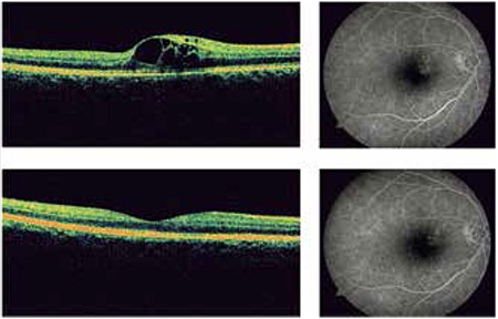
Six eyes of 5 patients with type 2 IMT who received intravitreal bevacizumab between 2009 and 2014 were included in this study. All the patients had an ophthalmological examination including best corrected visual acuity (BCVA), dilated fundus examination, spectral domain optical coherence tomography (OCT) and fluorescein angiography. Intravitreal bevacizumab injection was planned for patients who had macular edema and/or decreased visual acuity at baseline. Patients were examined 1 week and 1 month after the intravitreal injection. Intravitreal injection was repeated in patients whose visual acuity decreased and/or whose macular edema persisted or increased. Changes in BCVA, central macular thickness (CMT) and central macular volume from baseline at 1 month after the first injection and at final examination were evaluated.
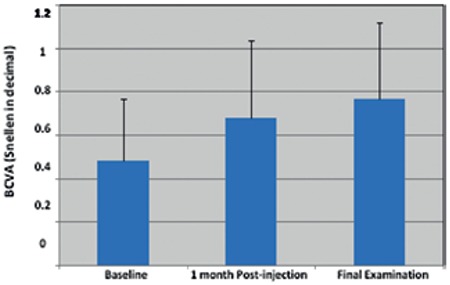
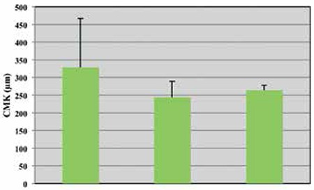
Average age of the patients (4 female and 1 male) was 62±11.8 years. Average follow-up period was 26±11 months. Patients received an average of 2.3 (range 1-4) injections during follow-up. Average Snellen BCVA of the patients was 0.48±0.29. BCVA increased at final examination compared to baseline in all of the patients. The difference between baseline and final visual acuities was significant (p<0.05). The patients' average CMT was 328±139 µm at baseline and decreased by a mean of 85±153 µm at 1 month after the first injection and 65±142 µm at final examination, but the changes were not significant. CMT decreased at final examination compared to baseline in four patients and increased in both eyes of one patient.
Intravitreal bevacizumab injection is a preferable treatment method in regard to both visual acuity and OCT findings.
评估玻璃体内注射贝伐单抗治疗2型特发性黄斑毛细血管扩张症(IMT)的疗效。
本研究纳入了2009年至2014年间接受玻璃体内注射贝伐单抗治疗的5例2型IMT患者的6只眼。所有患者均接受了眼科检查,包括最佳矫正视力(BCVA)、散瞳眼底检查、光谱域光学相干断层扫描(OCT)和荧光素血管造影。对于基线时存在黄斑水肿和/或视力下降的患者,计划进行玻璃体内注射贝伐单抗。在玻璃体内注射后1周和1个月对患者进行检查。对于视力下降和/或黄斑水肿持续或加重的患者,重复进行玻璃体内注射。评估首次注射后1个月及末次检查时与基线相比BCVA、中心黄斑厚度(CMT)和中心黄斑体积的变化。
患者(4例女性,1例男性)的平均年龄为62±11.8岁。平均随访时间为26±11个月。随访期间患者平均接受2.3次(范围1 - 4次)注射。患者的平均Snellen BCVA为0.48±0.29。与基线相比,所有患者在末次检查时BCVA均有所提高。基线和末次视力之间的差异具有统计学意义(p<0.05)。患者基线时平均CMT为328±139 µm,首次注射后1个月平均下降85±153 µm,末次检查时平均下降65±142 µm,但变化无统计学意义。与基线相比,4例患者在末次检查时CMT下降,1例患者双眼CMT增加。
就视力和OCT检查结果而言,玻璃体内注射贝伐单抗是一种较好的治疗方法。