Weiss Lucas Carolin, Tursunova Irada, Neuschmelting Volker, Nettekoven Charlotte, Oros-Peusquens Ana-Maria, Stoffels Gabriele, Faymonville Andrea Maria, Jon Shah N, Langen Karl Josef, Lockau Hannah, Goldbrunner Roland, Grefkes Christian
University of Cologne, Center of Neurosurgery, 50924 Cologne, Germany.
Institute of Neuroscience and Medicine, Research Centre Jülich, 52425 Jülich, Germany.
Neuroimage Clin. 2016 Nov 23;13:297-309. doi: 10.1016/j.nicl.2016.11.022. eCollection 2017.
DTI-based tractography is an increasingly important tool for planning brain surgery in patients suffering from brain tumours. However, there is an ongoing debate which tracking approaches yield the most valid results. Especially the use of functional localizer data such as navigated transcranial magnetic stimulation (nTMS) or functional magnetic resonance imaging (fMRI) seem to improve fibre tracking data in conditions where anatomical landmarks are less informative due to tumour-induced distortions of the gyral anatomy. We here compared which of the two localizer techniques yields more plausible results with respect to mapping different functional portions of the corticospinal tract (CST) in brain tumour patients.
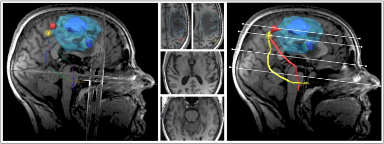
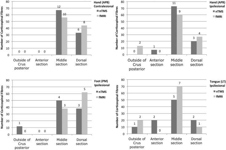
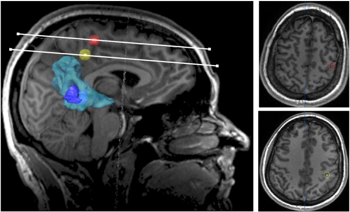
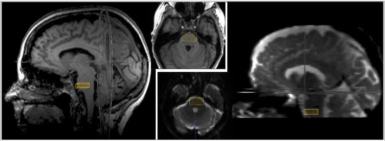
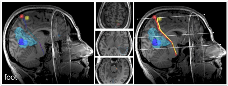
The CSTs of 18 patients with intracranial tumours in the vicinity of the primary motor area (M1) were investigated by means of deterministic DTI. The core zone of the tumour-adjacent hand, foot and/or tongue M1 representation served as cortical regions of interest (ROIs). M1 core zones were defined by both the nTMS hot-spots and the fMRI local activation maxima. In addition, for all patients, a subcortical ROI at the level of the inferior anterior pons was implemented into the tracking algorithm in order to improve the anatomical specificity of CST reconstructions. As intra-individual control, we additionally tracked the CST of the hand motor region of the unaffected, i.e., non-lesional hemisphere, again comparing fMRI and nTMS M1 seeds. The plausibility of the fMRI-ROI- vs. nTMS-ROI-based fibre trajectories was assessed by a-priori defined anatomical criteria. Moreover, the anatomical relationship of different fibre courses was compared regarding their distribution in the anterior-posterior direction as well as their location within the posterior limb of the internal capsule (PLIC).
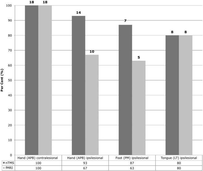
Overall, higher plausibility rates were observed for the use of nTMS- as compared to fMRI-defined cortical ROIs ( < 0.05) in tumour vicinity. On the non-lesional hemisphere, however, equally good plausibility rates (100%) were observed for both localizer techniques. fMRI-originated fibres generally followed a more posterior course relative to the nTMS-based tracts ( < 0.01) in both the lesional and non-lesional hemisphere.
NTMS achieved better tracking results than fMRI in conditions when the cortical tract origin (M1) was located in close vicinity to a brain tumour, probably influencing neurovascular coupling. Hence, especially in situations with altered BOLD signal physiology, nTMS seems to be the method of choice in order to identify seed regions for CST mapping in patients.
基于扩散张量成像(DTI)的纤维束成像对于脑肿瘤患者的脑外科手术规划而言,正成为一种日益重要的工具。然而,关于哪种追踪方法能产生最有效的结果,目前仍存在争议。特别是在因肿瘤导致脑回解剖结构变形,致使解剖标志信息不足的情况下,使用诸如导航经颅磁刺激(nTMS)或功能磁共振成像(fMRI)等功能定位数据,似乎能改善纤维追踪数据。我们在此比较了在脑肿瘤患者中,这两种定位技术中的哪一种在绘制皮质脊髓束(CST)的不同功能部分时能产生更合理的结果。
通过确定性DTI对18例颅内肿瘤位于初级运动区(M1)附近的患者的CST进行研究。肿瘤相邻的手部、足部和/或舌部M1代表区的核心区域作为皮质感兴趣区(ROI)。M1核心区域由nTMS热点和fMRI局部激活最大值共同定义。此外,对于所有患者,在追踪算法中加入脑桥前下部水平的皮质下ROI,以提高CST重建的解剖特异性。作为个体内对照,我们还追踪了未受影响即非病变半球的手部运动区的CST,同样比较了fMRI和nTMS的M1种子点。基于fMRI-ROI和基于nTMS-ROI的纤维轨迹的合理性通过预先定义的解剖学标准进行评估。此外,比较了不同纤维束在前后方向上的分布以及它们在内囊后肢(PLIC)内的位置的解剖学关系。
总体而言,在肿瘤附近,与基于fMRI定义皮质ROI相比,使用nTMS时观察到更高的合理性率(<0.05)。然而,在非病变半球,两种定位技术的合理性率均为100%。在病变半球和非病变半球中,相对于基于nTMS的纤维束,源自fMRI的纤维通常走行更靠后(<0.叭)。
当皮质纤维束起源(M1)紧邻脑肿瘤时,nTMS在追踪方面比fMRI取得了更好的结果,这可能影响了神经血管耦合。因此,特别是在血氧水平依赖(BOLD)信号生理改变的情况下,nTMS似乎是确定脑肿瘤患者CST映射种子区域的首选方法。