Chan C C, Ottesen E A, Awadzi K, Badu R, Nussenblatt R B
National Eye Institute, Bethesda, MD 20892.
Clin Exp Immunol. 1989 Sep;77(3):367-72.
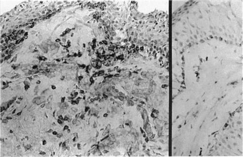
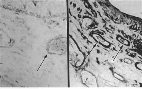
Ocular tissue (conjunctiva and iris) was obtained from 12 adult African men with active ocular onchocerciasis and from nine age-matched persons from the same endemic region but without onchocercal infection. These tissues were examined immunohistologically and two major findings were noted. First, mild-to-moderate chronic inflammatory cellular infiltration was present in the conjunctiva of the onchocerciasis patients. T lymphocytes (CD3+) were the major inflammatory cells, and the T suppressor/cytotoxic (CD8+) subset was significantly increased in the ocular onchocerciasis patients (P less than 0.03). Second, in the onchocerciasis patients, non-lymphoid cells of the conjunctiva and iris, such as vascular endothelium, pericytes and fibroblasts were in an activated state, as shown by increased expression of Class II MHC antigens (P less than 0.02, conjunctiva; P less than 0.05, iris). These concomitant findings of lymphocyte infiltration and resident cell activation indicate a dynamic state of localized host responsiveness presumably to the microfilarial parasites and their products in the anterior segments of the eyes of patients with ocular onchocerciasis.
从12名患有活动性眼部盘尾丝虫病的成年非洲男性以及9名来自同一流行地区、年龄匹配但无盘尾丝虫感染的人身上获取眼部组织(结膜和虹膜)。对这些组织进行了免疫组织学检查,发现了两个主要结果。首先,盘尾丝虫病患者的结膜存在轻度至中度慢性炎性细胞浸润。T淋巴细胞(CD3+)是主要的炎性细胞,眼部盘尾丝虫病患者的T抑制/细胞毒性(CD8+)亚群显著增加(P小于0.03)。其次,在盘尾丝虫病患者中,结膜和虹膜的非淋巴细胞,如血管内皮细胞、周细胞和成纤维细胞处于活化状态,这表现为II类MHC抗原表达增加(结膜P小于0.02;虹膜P小于0.05)。淋巴细胞浸润和驻留细胞活化的这些伴随发现表明,在眼部盘尾丝虫病患者的眼前节中,宿主对微丝蚴寄生虫及其产物可能存在局部反应的动态状态。