Shukla Shakti Dhar, Mahmood Malik Quasir, Weston Steven, Latham Roger, Muller Hans Konrad, Sohal Sukhwinder Singh, Walters Eugene Haydn
NHMRC Centre of Research Excellence for Chronic Respiratory Disease, School of Medicine, University of Tasmania, MS1, 17 Liverpool Street, Private Bag 23, Hobart, Tasmania, 7000, Australia.
School of Health Sciences, University of Tasmania, Launceston, Tasmania, 7248, Australia.
Respir Res. 2017 Jan 5;18(1):6. doi: 10.1186/s12931-016-0483-8.
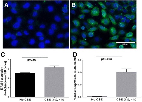
ICAM-1 is a major receptor for ~60% of human rhinoviruses, and non-typeable Haemophilus influenzae, two major pathogens in COPD. Increased cell-surface expression of ICAM-1 in response to tobacco smoke exposure has been suggested. We have investigated epithelial ICAM-1 expression in both the large and small airways, and lung parenchyma in smoking-related chronic airflow limitation (CAL) patients.
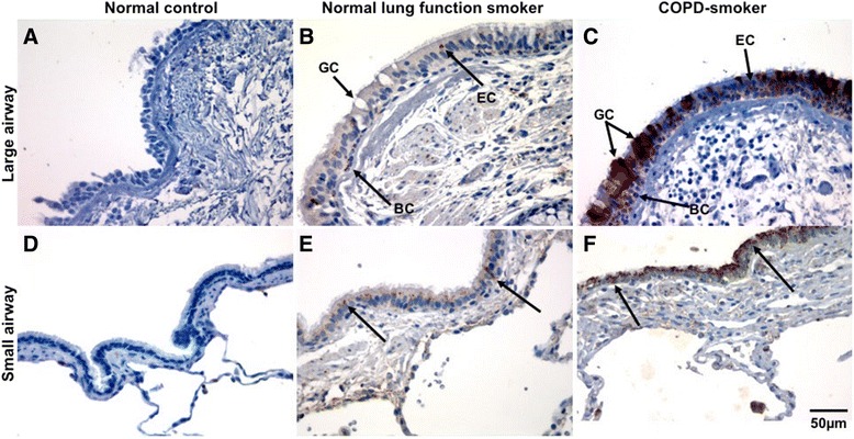
We evaluated epithelial ICAM-1 expression in resected lung tissue: 8 smokers with normal spirometry (NLFS); 29 CAL patients (10 small-airway disease; 9 COPD-smokers; 10 COPD ex-smokers); Controls (NC): 15 normal airway/lung tissues. Immunostaining with anti-ICAM-1 monoclonal antibody was quantified with computerized image analysis. The percent and type of cells expressing ICAM-1 in large and small airway epithelium and parenchyma were enumerated, plus percentage of epithelial goblet and submucosal glands positive for ICAM- 1.
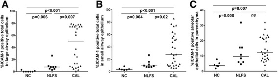
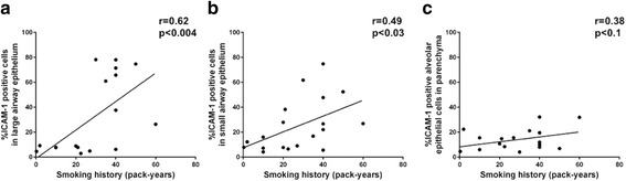
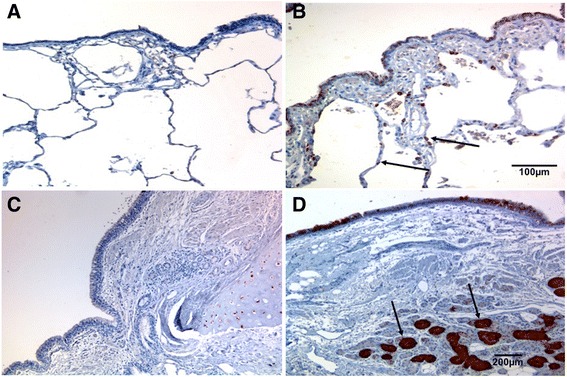
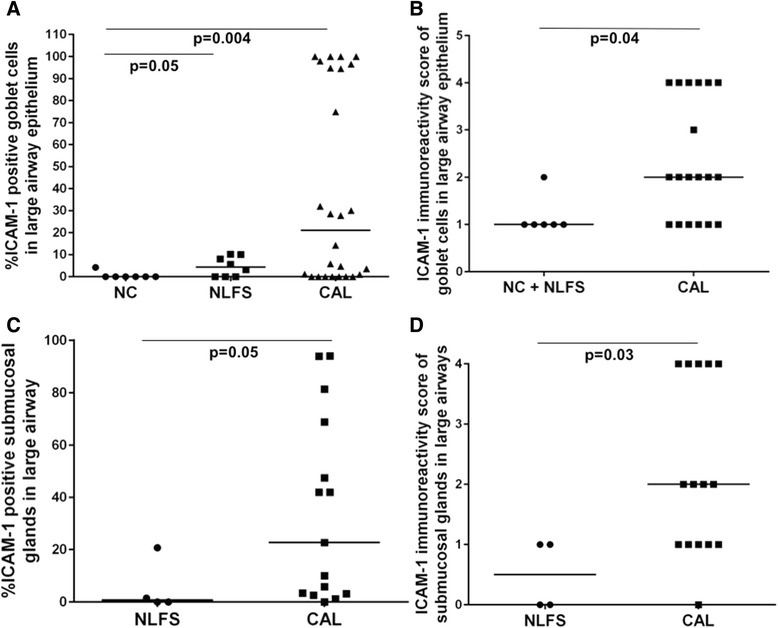
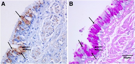
A major increase in ICAM-1 expression in epithelial cells was found in both large (p < 0.006) and small airways (p < 0.004) of CAL subjects compared to NC, with NLFS being intermediate. In the CAL group, both basal and luminal areas stained heavily for ICAM-1, so did goblet cells and sub-mucosal glands, however in either NC or NLFS subjects, only epithelial cell luminal surfaces stained. ICAM-1 expression on alveolar pneumocytes (mainly type II) was slightly increased in CAL and NLFS (p < 0.01). Pack-years of smoking correlated with ICAM-1 expression (r = 0.49; p < 0.03).
Airway ICAM-1 expression is markedly upregulated in CAL group, which could be crucial in rhinoviral and NTHi infections. The parenchymal ICAM-1 is affected by smoking, with no further enhancement in CAL subjects.
细胞间黏附分子-1(ICAM-1)是约60%的人鼻病毒和不可分型流感嗜血杆菌的主要受体,这两种是慢性阻塞性肺疾病(COPD)中的主要病原体。有研究表明,烟草烟雾暴露会导致ICAM-1在细胞表面的表达增加。我们研究了吸烟相关慢性气流受限(CAL)患者的大、小气道及肺实质中上皮细胞ICAM-1的表达情况。
我们评估了切除的肺组织中上皮细胞ICAM-1的表达:8名肺功能正常的吸烟者(NLFS);29名CAL患者(10名小气道疾病患者;9名吸烟的COPD患者;10名已戒烟的COPD患者);对照组(NC):15份正常气道/肺组织。使用抗ICAM-1单克隆抗体进行免疫染色,并通过计算机图像分析进行定量。统计大、小气道上皮及实质中表达ICAM-1的细胞百分比及类型,以及上皮杯状细胞和黏膜下腺中ICAM-1阳性的百分比。
与NC组相比,CAL组大(p < 0.006)、小气道(p < 0.004)上皮细胞中ICAM-1表达显著增加,NLFS组介于两者之间。在CAL组中,基底和管腔区域ICAM-1染色均较重,杯状细胞和黏膜下腺也是如此,但在NC组或NLFS组受试者中,仅上皮细胞管腔表面染色。CAL组和NLFS组肺泡上皮细胞(主要是II型)上ICAM-1的表达略有增加(p < 0.01)。吸烟包年数与ICAM-1表达相关(r = 0.49;p < 0.03)。
CAL组气道ICAM-1表达明显上调,这可能在鼻病毒和不可分型流感嗜血杆菌感染中起关键作用。肺实质ICAM-1受吸烟影响,在CAL患者中无进一步增强。