Henson John H, Ditzler Casey E, Germain Aphnie, Irwin Patrick M, Vogt Eric T, Yang Shucheng, Wu Xufeng, Shuster Charles B
Department of Biology, Dickinson College, Carlisle, PA 17013
Friday Harbor Laboratories, University of Washington, Friday Harbor, WA 98250.
Mol Biol Cell. 2017 Mar 1;28(5):613-623. doi: 10.1091/mbc.E16-06-0466. Epub 2017 Jan 5.
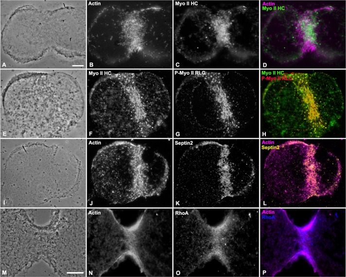
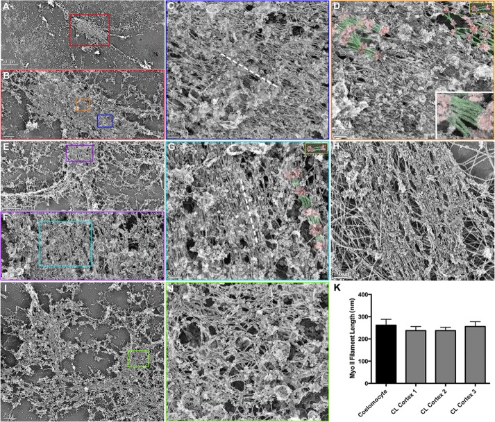
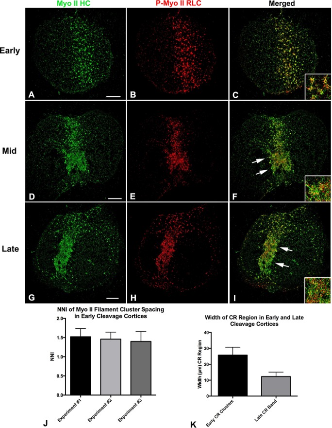
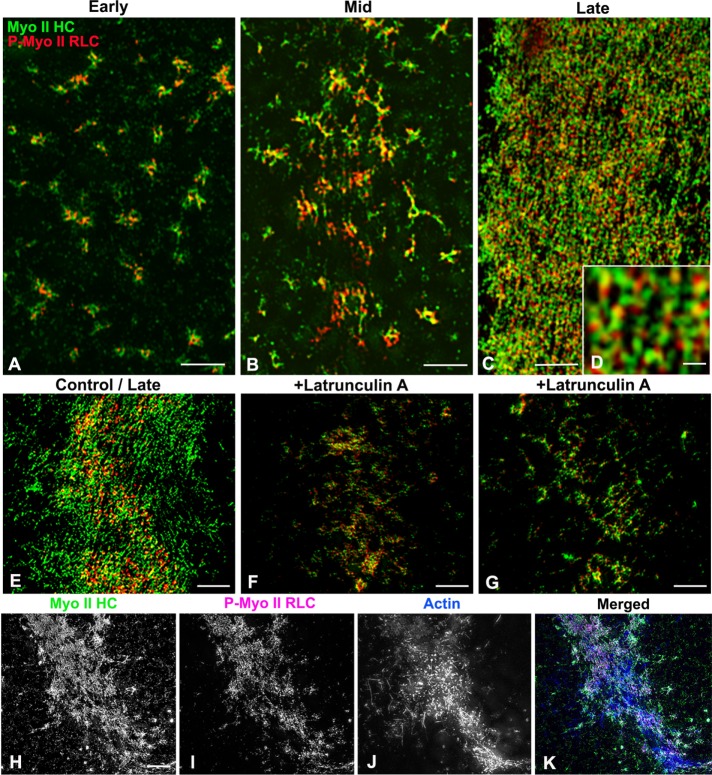
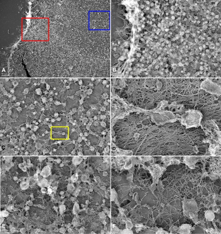
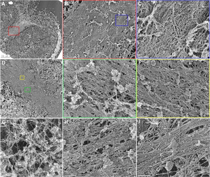
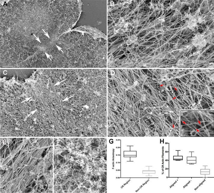
Despite recent advances in our understanding of the components and spatial regulation of the contractile ring (CR), the precise ultrastructure of actin and myosin II within the animal cell CR remains an unanswered question. We used superresolution light microscopy and platinum replica transmission electron microscopy (TEM) to determine the structural organization of actin and myosin II in isolated cortical cytoskeletons prepared from dividing sea urchin embryos. Three-dimensional structured illumination microscopy indicated that within the CR, actin and myosin II filaments were organized into tightly packed linear arrays oriented along the axis of constriction and restricted to a narrow zone within the furrow. In contrast, myosin II filaments in earlier stages of cytokinesis were organized into small, discrete, and regularly spaced clusters. TEM showed that actin within the CR formed a dense and anisotropic array of elongate, antiparallel filaments, whereas myosin II was organized into laterally associated, head-to-head filament chains highly reminiscent of mammalian cell stress fibers. Together these results not only support the canonical "purse-string" model for contractile ring constriction, but also suggest that the CR may be derived from foci of myosin II filaments in a manner similar to what has been demonstrated in fission yeast.
尽管我们对收缩环(CR)的组成成分和空间调控的理解最近取得了进展,但动物细胞CR内肌动蛋白和肌球蛋白II的确切超微结构仍是一个未解决的问题。我们使用超分辨率光学显微镜和铂复型透射电子显微镜(TEM)来确定从分裂的海胆胚胎制备的分离皮质细胞骨架中肌动蛋白和肌球蛋白II的结构组织。三维结构照明显微镜显示,在CR内,肌动蛋白和肌球蛋白II细丝被组织成紧密排列的线性阵列,沿收缩轴定向,并局限于沟内的一个狭窄区域。相比之下,胞质分裂早期的肌球蛋白II细丝被组织成小的、离散的且规则间隔的簇。TEM显示,CR内的肌动蛋白形成了密集且各向异性的细长、反平行细丝阵列,而肌球蛋白II则被组织成横向相关的、头对头的细丝链,这与哺乳动物细胞应力纤维非常相似。这些结果共同不仅支持收缩环收缩的经典“束带”模型,还表明CR可能以类似于裂殖酵母中所证明的方式从肌球蛋白II细丝的焦点衍生而来。