Farman H H, Wu J, Gustafsson K L, Windahl S H, Kim S H, Katzenellenbogen J A, Ohlsson C, Lagerquist M K
Centre for Bone and Arthritis ResearchInstitute of Medicine, Sahlgrenska Academy, University of Gothenburg, Gothenburg, Sweden
Centre for Bone and Arthritis ResearchInstitute of Medicine, Sahlgrenska Academy, University of Gothenburg, Gothenburg, Sweden.
J Mol Endocrinol. 2017 Feb;58(2):105-111. doi: 10.1530/JME-16-0209. Epub 2017 Jan 5.
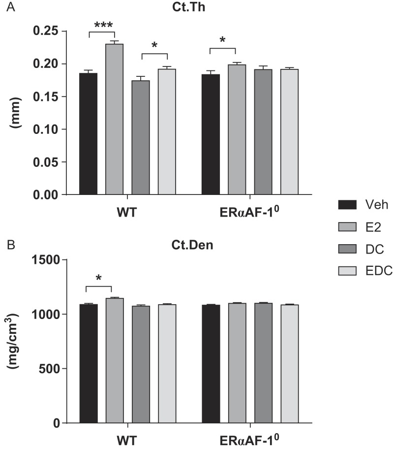
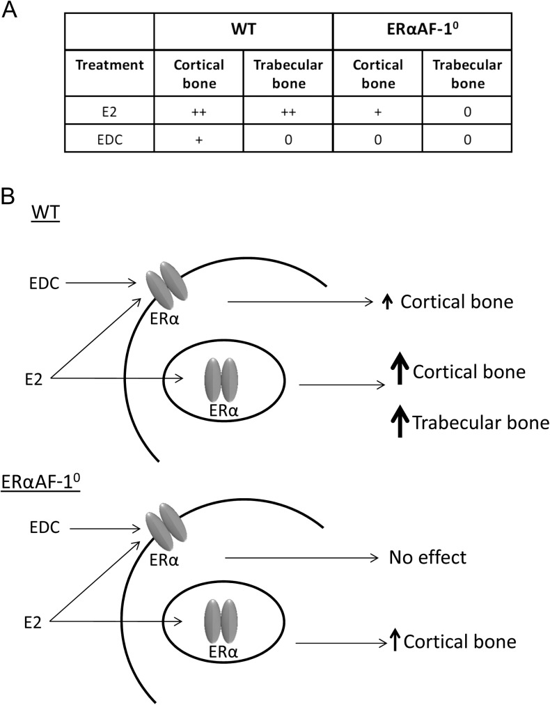
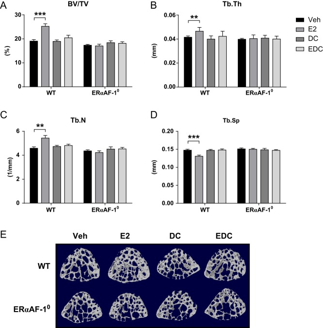
Estradiol (E2) signaling via estrogen receptor alpha (ERα) is important for the male skeleton as demonstrated by ERα inactivation in both mice and man. ERα mediates estrogenic effects not only by translocating to the nucleus and affecting gene transcription but also by extra-nuclear actions e.g., triggering cytoplasmic signaling cascades. ERα contains various domains, and the role of activation function 1 (ERαAF-1) is known to be tissue specific. The aim of this study was to determine the importance of extra-nuclear estrogen effects for the skeleton in males and to determine the role of ERαAF-1 for mediating these effects. Five-month-old male wild-type (WT) and ERαAF-1-inactivated (ERαAF-1) mice were orchidectomized and treated with equimolar doses of 17β-estradiol (E2) or an estrogen dendrimer conjugate (EDC), which is incapable of entering the nucleus and thereby only initiates extra-nuclear ER actions or their corresponding vehicles for 3.5 weeks. As expected, E2 treatment increased cortical thickness and trabecular bone volume per total volume (BV/TV) in WT males. EDC treatment increased cortical thickness in WT males, whereas no effect was detected in trabecular bone. In ERαAF-1 males, E2 treatment increased cortical thickness, but did not affect trabecular bone. Interestingly, the effect of EDC on cortical bone was abolished in ERαAF-1 mice. In conclusion, extra-nuclear estrogen signaling affects cortical bone mass in males, and this effect is dependent on a functional ERαAF-1. Increased knowledge regarding estrogen signaling mechanisms in the regulation of the male skeleton may aid the development of new treatment options for male osteoporosis.
通过雌激素受体α(ERα)的雌二醇(E2)信号传导对男性骨骼很重要,这已在小鼠和人类中通过ERα失活得到证实。ERα不仅通过转运到细胞核并影响基因转录来介导雌激素作用,还通过核外作用,例如触发细胞质信号级联反应。ERα包含多个结构域,已知激活功能1(ERαAF - 1)的作用具有组织特异性。本研究的目的是确定核外雌激素作用对男性骨骼的重要性,并确定ERαAF - 1在介导这些作用中的作用。对5个月大的雄性野生型(WT)和ERαAF - 1失活(ERαAF - 1)小鼠进行去势,并给予等摩尔剂量的17β - 雌二醇(E2)或雌激素树枝状聚合物缀合物(EDC)处理3.5周,EDC无法进入细胞核,因此仅引发核外ER作用或其相应的载体。正如预期的那样,E2处理增加了WT雄性小鼠的皮质厚度和每总体积的小梁骨体积(BV/TV)。EDC处理增加了WT雄性小鼠的皮质厚度,而在小梁骨中未检测到影响。在ERαAF - 1雄性小鼠中,E2处理增加了皮质厚度,但不影响小梁骨。有趣的是,在ERαAF - 1小鼠中,EDC对皮质骨的作用被消除。总之,核外雌激素信号传导影响男性的皮质骨量,并且这种作用依赖于功能性的ERαAF - 1。关于雌激素信号传导机制在男性骨骼调节中的知识增加可能有助于开发男性骨质疏松症的新治疗选择。