• 文献检索
  • 文档翻译
  • 深度研究
  • 学术资讯
  • Suppr Zotero 插件Zotero 插件
  • 邀请有礼
  • 套餐&价格
  • 历史记录
应用&插件
Suppr Zotero 插件Zotero 插件浏览器插件Mac 客户端Windows 客户端微信小程序
定价
高级版会员购买积分包购买API积分包
服务
文献检索文档翻译深度研究API 文档MCP 服务
关于我们
关于 Suppr公司介绍联系我们用户协议隐私条款
关注我们

Suppr 超能文献

核心技术专利:CN118964589B侵权必究
粤ICP备2023148730 号-1Suppr @ 2026

文献检索

告别复杂PubMed语法,用中文像聊天一样搜索,搜遍4000万医学文献。AI智能推荐,让科研检索更轻松。

立即免费搜索

文件翻译

保留排版,准确专业,支持PDF/Word/PPT等文件格式,支持 12+语言互译。

免费翻译文档

深度研究

AI帮你快速写综述,25分钟生成高质量综述,智能提取关键信息,辅助科研写作。

立即免费体验

氟脱氧葡萄糖正电子发射断层扫描-计算机断层扫描(FDG PET/CT)在一例表现为巨大颅外颅内肿块的罕见多发性骨髓瘤病例中的发现。

Fluorodeoxyglucose Positron Emission Tomography-Computed Tomography (FDG PET/CT) Findings in an Unusual Case of Multiple Myeloma Presenting with a Large Extra-Axial Intracranial Mass.

作者信息

Ayaz Sevin, Ayaz Ümit Yaşar

机构信息

Department of Nuclear Medicine, Mersin State Hospital, Mersin, Turkey.

Department of Radiology, Mersin Women's and Children's Hospital, Mersin, Turkey.

出版信息

Pol J Radiol. 2016 Dec 16;81:602-605. doi: 10.12659/PJR.899444. eCollection 2016.

DOI:10.12659/PJR.899444
PMID:28058074
原文链接:https://pmc.ncbi.nlm.nih.gov/articles/PMC5175789/
Abstract

BACKGROUND

We aimed to present unusual cranial FDG PET/CT findings of a 56-year-old female with multiple myeloma (MM).

CASE REPORT

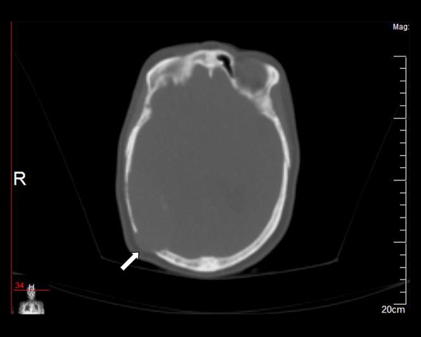
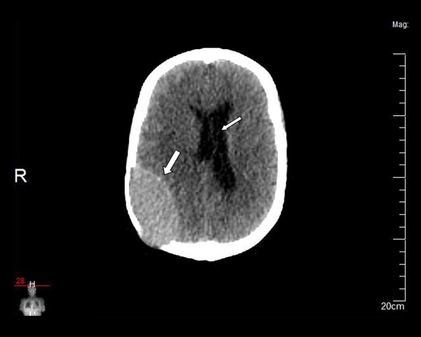
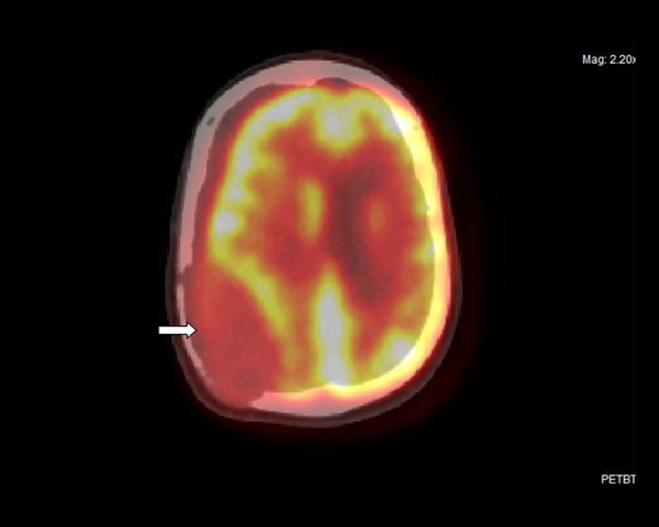
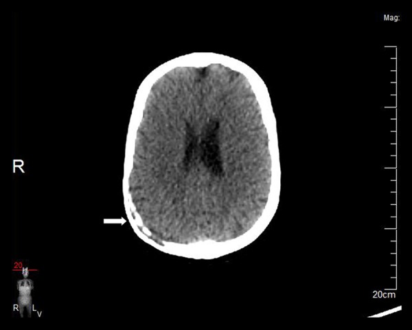
Plain CT images revealed a lytic lesion in the right parietal bone, filled with an oval-shaped, large, extra-axial, extradural, intracranial mass which measured 75×75×40 mm and had smooth borders. The right parietal lobe was compressed by the mass. The maximum standardized uptake value (SUV) of the mass lesion was 8.94 on FDG PET/CT images. Multiple lytic lesions with an increased uptake were also detected in other calvarial bones, in several vertebras and in the proximal left femur. After seven months, a control FDG PET/CT following radiotherapy and chemotherapy revealed almost complete regression of the right parietal extra-axial mass lesion. The number, size and metabolism of lytic lesions in other bones also decreased.

CONCLUSIONS

FDG PET/CT was useful for an initial evaluation of MM lesions and was effective in monitoring the response of these lesions to therapy.

摘要

背景

我们旨在展示一名56岁多发性骨髓瘤(MM)女性患者不寻常的颅骨FDG PET/CT表现。

病例报告

平扫CT图像显示右侧顶骨有一个溶骨性病变,内有一个椭圆形、巨大、轴外、硬膜外、颅内肿块,大小为75×75×40 mm,边界光滑。右侧顶叶被肿块压迫。在FDG PET/CT图像上,肿块病变的最大标准化摄取值(SUV)为8.94。在其他颅骨、多个椎体和左股骨近端也检测到多个溶骨性病变且摄取增加。七个月后,放疗和化疗后的对照FDG PET/CT显示右侧顶骨轴外肿块病变几乎完全消退。其他骨骼中溶骨性病变的数量、大小和代谢也有所减少。

结论

FDG PET/CT对MM病变的初始评估有用,并且在监测这些病变对治疗的反应方面有效。

https://cdn.ncbi.nlm.nih.gov/pmc/blobs/e3df/5175789/f22ab5be5cf1/poljradiol-81-602-g005.jpg
https://cdn.ncbi.nlm.nih.gov/pmc/blobs/e3df/5175789/2473a9813a92/poljradiol-81-602-g001.jpg
https://cdn.ncbi.nlm.nih.gov/pmc/blobs/e3df/5175789/486b74eb5b89/poljradiol-81-602-g002.jpg
https://cdn.ncbi.nlm.nih.gov/pmc/blobs/e3df/5175789/faaa92a29aa2/poljradiol-81-602-g003.jpg
https://cdn.ncbi.nlm.nih.gov/pmc/blobs/e3df/5175789/8f5070f25852/poljradiol-81-602-g004.jpg
https://cdn.ncbi.nlm.nih.gov/pmc/blobs/e3df/5175789/f22ab5be5cf1/poljradiol-81-602-g005.jpg

相似文献

1
Fluorodeoxyglucose Positron Emission Tomography-Computed Tomography (FDG PET/CT) Findings in an Unusual Case of Multiple Myeloma Presenting with a Large Extra-Axial Intracranial Mass.氟脱氧葡萄糖正电子发射断层扫描-计算机断层扫描(FDG PET/CT)在一例表现为巨大颅外颅内肿块的罕见多发性骨髓瘤病例中的发现。
Pol J Radiol. 2016 Dec 16;81:602-605. doi: 10.12659/PJR.899444. eCollection 2016.
2
More advantages in detecting bone and soft tissue metastases from prostate cancer using F-PSMA PET/CT.使用F-PSMA PET/CT检测前列腺癌骨和软组织转移方面有更多优势。
Hell J Nucl Med. 2019 Jan-Apr;22(1):6-9. doi: 10.1967/s002449910952. Epub 2019 Mar 7.
3
(18)F-FDG positron emission tomography/computed tomography and (99m)Tc-MDP skeletal scintigraphy in a case of Erdheim-Chester disease.18F-氟代脱氧葡萄糖正电子发射断层扫描/计算机断层扫描及99m锝-亚甲基二膦酸盐骨闪烁显像在1例厄尔海姆-切斯特病中的应用
Hell J Nucl Med. 2011 Sep-Dec;14(3):311-2.
4
Fluorodeoxyglucose positron emission tomography with computed tomography detects greater metabolic changes that are not represented by plain radiography for patients with osteonecrosis of the jaw.对于颌骨坏死患者,氟脱氧葡萄糖正电子发射断层扫描联合计算机断层扫描能检测到更大的代谢变化,而这些变化在X线平片中无法显示。
J Oral Maxillofac Surg. 2014 Oct;72(10):1957-65. doi: 10.1016/j.joms.2014.04.017. Epub 2014 Apr 24.
5
Different lesions revealed by 18F-FDG PET/CT and 18F-NaF PET/CT in patients with multiple myeloma.18F-FDG PET/CT 和 18F-NaF PET/CT 显示多发性骨髓瘤患者的不同病变。
Clin Nucl Med. 2014 Sep;39(9):e407-9. doi: 10.1097/RLU.0000000000000285.
6
Application of PET/CT in treatment response evaluation and recurrence prediction in patients with newly-diagnosed multiple myeloma.PET/CT在新诊断多发性骨髓瘤患者治疗反应评估及复发预测中的应用
Oncotarget. 2017 Apr 11;8(15):25637-25649. doi: 10.18632/oncotarget.11418.
7
Correlation Between Baseline 18F-FDG PET/CT Findings and CD38- and CD138-Expressing Myeloma Cells in Bone Marrow and Clinical Parameters in Patients with Multiple Myeloma.多发性骨髓瘤患者基线18F-FDG PET/CT检查结果与骨髓中表达CD38和CD138的骨髓瘤细胞及临床参数之间的相关性
Turk J Haematol. 2018 Aug 3;35(3):175-180. doi: 10.4274/tjh.2017.0372. Epub 2018 May 28.
8
11C-choline vs. 18F-FDG PET/CT in assessing bone involvement in patients with multiple myeloma.11C-胆碱与18F-氟代脱氧葡萄糖PET/CT在评估多发性骨髓瘤患者骨受累情况中的比较
World J Surg Oncol. 2007 Jun 20;5:68. doi: 10.1186/1477-7819-5-68.
9
Case Report: A rare case of multicentric angiosarcomas of bone mimicking multiple myeloma on F-FDG PET/CT.病例报告:1例罕见的骨多中心血管肉瘤在F-FDG PET/CT上酷似多发性骨髓瘤。
Front Med (Lausanne). 2023 Nov 28;10:1330341. doi: 10.3389/fmed.2023.1330341. eCollection 2023.
10
Semiquantitative assessment of osteoblastic, osteolytic, and mixed lytic-sclerotic bone lesions on fluorodeoxyglucose positron emission tomography/computed tomography and bone scintigraphy.氟脱氧葡萄糖正电子发射断层扫描/计算机断层扫描及骨闪烁显像上成骨性、溶骨性和混合性溶骨-硬化性骨病变的半定量评估
World J Nucl Med. 2019 Apr-Jun;18(2):132-136. doi: 10.4103/wjnm.WJNM_31_18.

引用本文的文献

1
Effects of Histone Deacetylase Inhibitor Panobinostat (LBH589) on Bone Marrow Mononuclear Cells of Relapsed or Refractory Multiple Myeloma Patients and Its Mechanisms.组蛋白去乙酰化酶抑制剂帕比司他(LBH589)对复发或难治性多发性骨髓瘤患者骨髓单个核细胞的影响及其机制。
Med Sci Monit. 2017 Oct 29;23:5150-5157. doi: 10.12659/msm.904232.

本文引用的文献

1
Calvarial tuberculosis presenting as cystic lesion: An unusual presentation in two patients.表现为囊性病变的颅骨结核:两例不寻常病例报告
J Cytol. 2015 Jul-Sep;32(3):176-80. doi: 10.4103/0970-9371.168844.
2
Insufficiency of bone scintigraphy in vertebral lesions of langerhans cell histiocytosis compared to f-18 fluorodeoxyglucose positron emission tomography/computed tomography and diagnostic computed tomography.与F-18氟脱氧葡萄糖正电子发射断层扫描/计算机断层扫描及诊断性计算机断层扫描相比,骨闪烁扫描在朗格汉斯细胞组织细胞增多症椎体病变中的不足
Mol Imaging Radionucl Ther. 2015 Feb 5;24(1):21-4. doi: 10.4274/mirt.58066.
3
False Positive F-18 FDG PET/CT of Skeletal Metastasis Due to Solitary Eosinophilic Granuloma.
孤立性嗜酸性肉芽肿导致骨骼转移的 F-18 FDG PET/CT 假阳性
Mol Imaging Radionucl Ther. 2013 Dec;22(3):103-5. doi: 10.4274/Mirt.296. Epub 2013 Dec 10.
4
Evolving role of FDG PET/CT in multiple myeloma imaging and management.18F-FDG PET/CT 在多发性骨髓瘤影像学诊断及治疗中的作用演变。
AJR Am J Roentgenol. 2013 Apr;200(4):884-90. doi: 10.2214/AJR.12.9653.
5
Multiple myeloma imaging.
Diagn Interv Imaging. 2013 Feb;94(2):190-2. doi: 10.1016/j.diii.2012.12.003. Epub 2013 Jan 16.
6
Positron emission tomography/computed tomography imaging of brown tumors mimicking multiple skeletal metastases in patient with primary hyperparathyroidism.原发性甲状旁腺功能亢进患者中,棕色瘤正电子发射断层扫描/计算机断层扫描成像表现酷似多发性骨转移。
Indian J Endocrinol Metab. 2012 Sep;16(5):850-2. doi: 10.4103/2230-8210.100682.
7
Cancer statistics, 2011: the impact of eliminating socioeconomic and racial disparities on premature cancer deaths.癌症统计数据,2011 年:消除社会经济和种族差异对癌症过早死亡的影响。
CA Cancer J Clin. 2011 Jul-Aug;61(4):212-36. doi: 10.3322/caac.20121. Epub 2011 Jun 17.
8
[Calvarial tuberculosis: two case reports].
Arch Pediatr. 2011 Apr;18(4):397-400. doi: 10.1016/j.arcped.2011.01.026.
9
Multiple myeloma presenting as an intracranial plasmacytoma: a case report.以颅内浆细胞瘤形式出现的多发性骨髓瘤:一例报告
Cases J. 2009 Nov 30;2:9110. doi: 10.1186/1757-1626-2-9110.
10
Role of radiography, MRI and FDG-PET/CT in diagnosing, staging and therapeutical evaluation of patients with multiple myeloma.X线摄影、磁共振成像及氟代脱氧葡萄糖正电子发射断层显像/计算机断层扫描在多发性骨髓瘤患者诊断、分期及治疗评估中的作用
Ann Hematol. 2009 Dec;88(12):1161-8. doi: 10.1007/s00277-009-0829-0.