Byrd Darrin W, Doot Robert K, Allberg Keith C, MacDonald Lawrence R, McDougald Wendy A, Elston Brian F, Linden Hannah M, Kinahan Paul E
Department of Radiology, University of Washington, Seattle, Washington.
Department of Radiology, University of Pennsylvania, Philadelphia, Pennsylvania.
Tomography. 2016 Dec;2(4):353-360. doi: 10.18383/j.tom.2016.00205.
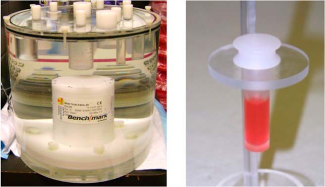
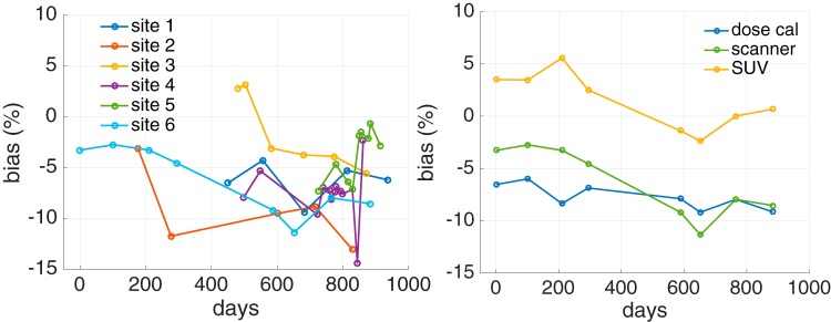
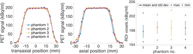
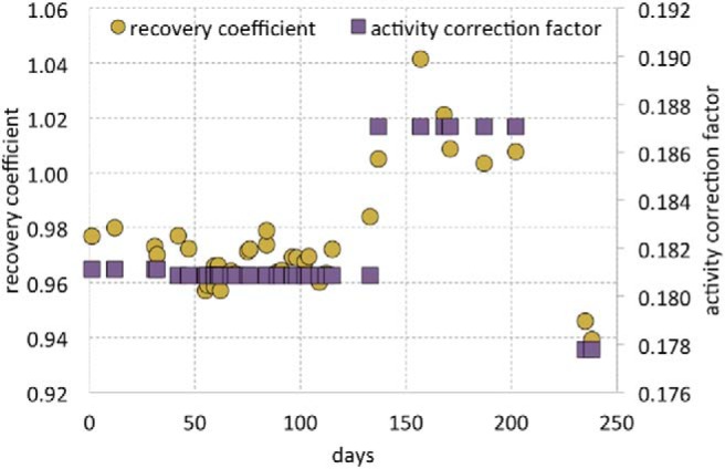
Quantitative PET imaging is an important tool for clinical trials evaluating the response of cancers to investigational therapies. The standardized uptake value, used as a quantitative imaging biomarker, is dependent on multiple parameters that may contribute bias and variability. The use of long-lived, sealed PET calibration phantoms offers the advantages of known radioactivity activity concentration and simpler use than aqueous phantoms. We evaluated scanner and dose calibrator sources from two batches of commercially available kits, together at a single site and distributed across a local multicenter PET imaging network. We found that radioactivity concentration was uniform within the phantoms. Within the regions of interest drawn in the phantom images, coefficients of variation of voxel values were less than 2%. Across phantoms, coefficients of variation for mean signal were close to 1%. Biases of the standardized uptake value estimated with the kits varied by site and were seen to change in time by approximately ±5%. We conclude that these biases cannot be assumed constant over time. The kits provide a robust method to monitor PET scanner and dose calibrator biases, and resulting biases in standardized uptake values.
定量PET成像是评估癌症对研究性疗法反应的临床试验的重要工具。用作定量成像生物标志物的标准化摄取值取决于多个可能导致偏差和变异性的参数。使用长寿命、密封的PET校准体模具有放射性活度浓度已知且比水溶液体模使用更简便的优点。我们在单个站点共同评估了来自两批市售试剂盒的扫描仪和剂量校准源,并将其分布在当地的多中心PET成像网络中。我们发现体模内的放射性浓度是均匀的。在体模图像中绘制的感兴趣区域内,体素值的变异系数小于2%。在不同体模中,平均信号的变异系数接近1%。用试剂盒估计的标准化摄取值的偏差因站点而异,并且随时间变化约±5%。我们得出结论,不能假定这些偏差随时间恒定。这些试剂盒提供了一种可靠的方法来监测PET扫描仪和剂量校准器的偏差以及标准化摄取值中产生的偏差。