• 文献检索
  • 文档翻译
  • 深度研究
  • 学术资讯
  • Suppr Zotero 插件Zotero 插件
  • 邀请有礼
  • 套餐&价格
  • 历史记录
应用&插件
Suppr Zotero 插件Zotero 插件浏览器插件Mac 客户端Windows 客户端微信小程序
定价
高级版会员购买积分包购买API积分包
服务
文献检索文档翻译深度研究API 文档MCP 服务
关于我们
关于 Suppr公司介绍联系我们用户协议隐私条款
关注我们

Suppr 超能文献

核心技术专利:CN118964589B侵权必究
粤ICP备2023148730 号-1Suppr @ 2026

文献检索

告别复杂PubMed语法,用中文像聊天一样搜索,搜遍4000万医学文献。AI智能推荐,让科研检索更轻松。

立即免费搜索

文件翻译

保留排版,准确专业,支持PDF/Word/PPT等文件格式,支持 12+语言互译。

免费翻译文档

深度研究

AI帮你快速写综述,25分钟生成高质量综述,智能提取关键信息,辅助科研写作。

立即免费体验

低级别基底动脉粥样硬化的斑块分布及其临床意义。

Plaque distribution of low-grade basilar artery atherosclerosis and its clinical relevance.

作者信息

Yu Jin, Li Ming-Li, Xu Yu-Yuan, Wu Shi-Wen, Lou Min, Mu Xue-Tao, Feng Feng, Gao Shan, Xu Wei-Hai

机构信息

Department of Neurology and Radiology, Peking Union Medical College Hospital, Chinese Academy of Medical Sciences, Shuaifuyuan1, Dongcheng District, Beijing, 100730, China.

Department of Neurology and Radiology, General Hospital of Chinese People's Armed Police Force, Beijing, China.

出版信息

BMC Neurol. 2017 Jan 10;17(1):8. doi: 10.1186/s12883-016-0785-y.

DOI:10.1186/s12883-016-0785-y
PMID:28068949
原文链接:https://pmc.ncbi.nlm.nih.gov/articles/PMC5223551/
Abstract

BACKGROUND

The underlying pathophysiology of BA distribution is unclear and intriguing. Using high-resolution magnetic resonance imaging (HR-MRI), we sought to explore the plaque distribution of low-grade basilar artery (BA) atherosclerosis and its clinical relevance.

METHODS

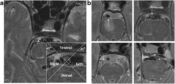
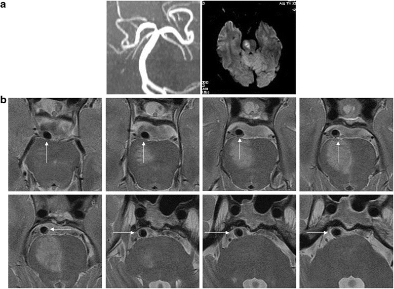
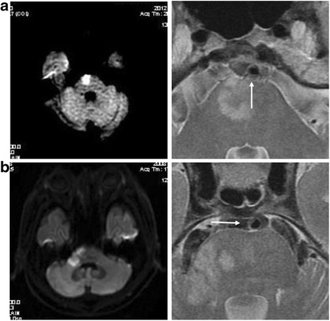
We retrospectively analyzed the imaging and clinical data of 61 patients with low-grade atherosclerotic BA stenosis (<50%). On HR-MRI, the plaques were categorized based on the involvement of the ventral, dorsal, or lateral sides of BA wall. A culprit plaque was defined if it was on the same slice or neighboring slices of symptomatic pontine infarcts and played a probable causal role (dorsal plaques with median pontine infarcts or lateral plaques with ipsilateral pontine infarcts). The relationships between plaque distribution and clinical presentations were analyzed.

RESULTS

Twenty-five symptomatic and thirty-six asymptomatic BAs with 752 HR-MRI image slices were studied. The average length of BA atherosclerosis plaques was 12.16 ± 5.61mm (10.30 ± 6.44mm in symptomatic and 13.46 ± 7.03mm in asymptomatic patients, p = 0.079). The plaque distribution was similar at ventral (29.0%), dorsal (37.6%) and lateral walls (33.1%). The BA plaques in symptomatic patients were more frequently located at the dorsal (42.5%) and lateral (41.2%) walls than at the ventral walls (16.1%; P < 0.05). Compared with symptomatic patients, asymptomatic patients more likely had their plaques distributed at the ventral walls (P = 0.022). Culprit plaques were observed in 85.0% (17/20) pontine infarcts in symptomatic patients and only 14.3% (2/14) silent pontine infarcts in asymptomatic patients (p < 0.001).

CONCLUSIONS

Low-grade BA atherosclerosis has a long distribution and evenly involves ventral, dorsal and lateral walls. The plaques at dorsal and lateral walls are associated with symptomatic pontine infarcts but not with silent infarcts.

摘要

背景

基底动脉(BA)粥样硬化斑块分布的潜在病理生理学尚不清楚且引人关注。我们使用高分辨率磁共振成像(HR-MRI)来探究轻度基底动脉粥样硬化的斑块分布及其临床相关性。

方法

我们回顾性分析了61例轻度动脉粥样硬化性BA狭窄(<50%)患者的影像学和临床资料。在HR-MRI上,根据斑块累及BA壁的腹侧、背侧或外侧对斑块进行分类。如果斑块位于有症状脑桥梗死的同一层面或相邻层面且可能起因果作用(背侧斑块导致脑桥中部梗死或外侧斑块导致同侧脑桥梗死),则定义为责任斑块。分析斑块分布与临床表现之间的关系。

结果

对25条有症状和36条无症状的BA的752个HR-MRI图像层面进行了研究。BA动脉粥样硬化斑块的平均长度为12.16±5.61mm(有症状患者为10.30±6.44mm,无症状患者为13.46±7.03mm,p = 0.079)。斑块在腹侧(29.0%)、背侧(37.6%)和外侧壁(33.1%)的分布相似。有症状患者的BA斑块更多位于背侧(42.5%)和外侧壁(41.2%),而非腹侧壁(16.1%;P<0.05)。与有症状患者相比,无症状患者的斑块更可能分布在腹侧壁(P = 0.022)。在有症状患者中,85.0%(17/20)的脑桥梗死存在责任斑块,而在无症状患者中,只有14.3%(2/14)的无症状脑桥梗死存在责任斑块(p<0.001)。

结论

轻度BA粥样硬化有较长的分布范围,且腹侧、背侧和外侧壁均有累及。背侧和外侧壁的斑块与有症状的脑桥梗死相关,但与无症状梗死无关。

https://cdn.ncbi.nlm.nih.gov/pmc/blobs/3160/5223551/f002161cae36/12883_2016_785_Fig3_HTML.jpg
https://cdn.ncbi.nlm.nih.gov/pmc/blobs/3160/5223551/42d5ae45a36b/12883_2016_785_Fig1_HTML.jpg
https://cdn.ncbi.nlm.nih.gov/pmc/blobs/3160/5223551/ac937a3d1966/12883_2016_785_Fig2_HTML.jpg
https://cdn.ncbi.nlm.nih.gov/pmc/blobs/3160/5223551/f002161cae36/12883_2016_785_Fig3_HTML.jpg
https://cdn.ncbi.nlm.nih.gov/pmc/blobs/3160/5223551/42d5ae45a36b/12883_2016_785_Fig1_HTML.jpg
https://cdn.ncbi.nlm.nih.gov/pmc/blobs/3160/5223551/ac937a3d1966/12883_2016_785_Fig2_HTML.jpg
https://cdn.ncbi.nlm.nih.gov/pmc/blobs/3160/5223551/f002161cae36/12883_2016_785_Fig3_HTML.jpg

相似文献

1
Plaque distribution of low-grade basilar artery atherosclerosis and its clinical relevance.低级别基底动脉粥样硬化的斑块分布及其临床意义。
BMC Neurol. 2017 Jan 10;17(1):8. doi: 10.1186/s12883-016-0785-y.
2
Relationship between the geometry patterns of vertebrobasilar artery and atherosclerosis.椎基底动脉几何形态与动脉粥样硬化之间的关系。
BMC Neurol. 2018 Jun 12;18(1):83. doi: 10.1186/s12883-018-1084-6.
3
Etiology of isolated pontine infarctions: a study based on high-resolution MRI and brain small vessel disease scores.孤立性脑桥梗死的病因学:一项基于高分辨率MRI和脑小血管病评分的研究
BMC Neurol. 2017 Dec 12;17(1):216. doi: 10.1186/s12883-017-0999-7.
4
Evaluation of basilar artery atherosclerotic plaque distribution by 3D MR vessel wall imaging.通过三维磁共振血管壁成像评估基底动脉粥样硬化斑块分布
J Magn Reson Imaging. 2016 Dec;44(6):1592-1599. doi: 10.1002/jmri.25296. Epub 2016 Jun 1.
5
Visualization of basilar artery atherosclerotic plaques by conventional T2-weighted magnetic resonance imaging: A case-control study.常规 T2 加权磁共振成像对基底动脉粥样硬化斑块的可视化:一项病例对照研究。
PLoS One. 2019 Feb 26;14(2):e0212570. doi: 10.1371/journal.pone.0212570. eCollection 2019.
6
Incremental Value of Plaque Enhancement in Patients with Moderate or Severe Basilar Artery Stenosis: 3.0 T High-Resolution Magnetic Resonance Study.中重度基底动脉狭窄患者斑块强化的增量价值:3.0T 高分辨率磁共振研究。
Biomed Res Int. 2017;2017:4281629. doi: 10.1155/2017/4281629. Epub 2017 Sep 17.
7
Basilar artery atherosclerotic plaques distribution in symptomatic patients: a 3.0T high-resolution MRI study.症状性患者基底动脉粥样硬化斑块分布:3.0T 高分辨率 MRI 研究。
Eur J Radiol. 2013 Apr;82(4):e199-203. doi: 10.1016/j.ejrad.2012.10.031. Epub 2012 Dec 8.
8
Plaques of Nonstenotic Basilar Arteries with Isolated Pontine Infarction on Three-dimensional High Isotropic Resolution Magnetic Resonance Imaging.三维高各向同性分辨率磁共振成像显示非狭窄性基底动脉斑块伴孤立性脑桥梗死
Chin Med J (Engl). 2015 Jun 5;128(11):1433-7. doi: 10.4103/0366-6999.157633.
9
Basilar artery atherosclerotic plaques in paramedian and lacunar pontine infarctions: a high-resolution MRI study.基底动脉粥样硬化斑块在旁正中及腔隙性脑桥梗死中的作用:高分辨率 MRI 研究。
Stroke. 2010 Jul;41(7):1405-9. doi: 10.1161/STROKEAHA.110.583534. Epub 2010 Jun 10.
10
Vessel wall differences between middle cerebral artery and basilar artery plaques on magnetic resonance imaging.磁共振成像显示大脑中动脉和基底动脉斑块的血管壁差异。
Sci Rep. 2016 Dec 5;6:38534. doi: 10.1038/srep38534.

引用本文的文献

1
Evaluation of the effect on stroke mechanism, stroke recurrence and clinical outcome in stroke patients with basilar artery atherosclerosis: A single centre retrospective observational study.评价基底动脉粥样硬化性脑卒中患者的卒中机制、卒中复发和临床结局的影响:一项单中心回顾性观察研究。
Sci Prog. 2024 Oct-Dec;107(4):368504241301519. doi: 10.1177/00368504241301519.
2
Preliminary results on temporal evolution and clinical implications of atherosclerotic plaque in branch atheromatous disease after statin treatment.他汀治疗后分支动脉粥样硬化疾病中动脉粥样硬化斑块的时间演变及临床意义的初步结果。
Ther Adv Neurol Disord. 2024 Sep 18;17:17562864241273902. doi: 10.1177/17562864241273902. eCollection 2024.
3

本文引用的文献

1
High-resolution MRI of intracranial atherosclerotic disease.颅内动脉粥样硬化疾病的高分辨率磁共振成像
Neurointervention. 2014 Feb;9(1):9-20. doi: 10.5469/neuroint.2014.9.1.9. Epub 2014 Feb 28.
2
Basilar artery atherosclerosis and hypertensive small vessel disease in isolated pontine infarctions: a study based on high-resolution MRI.孤立性脑桥梗死中基底动脉粥样硬化与高血压小血管病:基于高分辨率 MRI 的研究。
Eur Neurol. 2013;70(1-2):16-21. doi: 10.1159/000346577. Epub 2013 May 7.
3
Basilar artery atherosclerotic plaques distribution in symptomatic patients: a 3.0T high-resolution MRI study.
Hemodynamic characteristics of vertebrobasilar artery fenestration combined with vertebrobasilar dolichoectasia: a study based on magnetic resonance angiography.
椎动脉基底动脉开窗合并椎动脉基底动脉延长扩张症的血流动力学特征:一项基于磁共振血管造影的研究
Am J Nucl Med Mol Imaging. 2024 Aug 25;14(4):253-260. doi: 10.62347/TLNN8316. eCollection 2024.
4
Risk prediction of CISS classification in endovascular treatment of basilar artery stenosis.基底动脉狭窄血管内治疗中CISS分型的风险预测
Heliyon. 2023 Dec 16;10(1):e23747. doi: 10.1016/j.heliyon.2023.e23747. eCollection 2024 Jan 15.
5
Effect of dominant vertebral artery angle on basilar artery curvature and plaque.优势椎动脉角度对基底动脉曲率和斑块的影响。
Quant Imaging Med Surg. 2023 Sep 1;13(9):5748-5758. doi: 10.21037/qims-23-74. Epub 2023 Jul 10.
6
Patterns and implications of artery remodeling based on high-resolution vessel wall imaging in symptomatic severe basilar artery stenosis.基于高分辨率血管壁成像的有症状严重基底动脉狭窄的动脉重塑模式及影响
Quant Imaging Med Surg. 2023 Apr 1;13(4):2098-2108. doi: 10.21037/qims-22-771. Epub 2023 Feb 6.
7
Clinical Relevance of Plaque Distribution for Basilar Artery Stenosis.基底动脉狭窄斑块分布的临床相关性。
AJNR Am J Neuroradiol. 2023 May;44(5):530-535. doi: 10.3174/ajnr.A7839. Epub 2023 Apr 6.
8
Basilar artery plaque distribution is associated with pontine infarction and vertebrobasilar artery geometry.基底动脉斑块分布与脑桥梗死及椎基底动脉形态有关。
Front Neurol. 2023 Mar 13;14:1079905. doi: 10.3389/fneur.2023.1079905. eCollection 2023.
9
Association between basilar artery stenosis features, vertebral artery stenosis and perforator stroke after stenting.支架置入术后基底动脉狭窄特征、椎动脉狭窄与穿支卒中之间的关联。
Interv Neuroradiol. 2025 Apr;31(2):246-254. doi: 10.1177/15910199231164840. Epub 2023 Mar 29.
10
Relationship Between Hypertension and Basilar Atherosclerosis in Chinese Han Population: A High-Resolution Magnetic Resonance Imaging Study.中国汉族人群高血压与基底动脉粥样硬化的关系:一项高分辨率磁共振成像研究
Front Cardiovasc Med. 2022 Apr 27;9:830664. doi: 10.3389/fcvm.2022.830664. eCollection 2022.
症状性患者基底动脉粥样硬化斑块分布:3.0T 高分辨率 MRI 研究。
Eur J Radiol. 2013 Apr;82(4):e199-203. doi: 10.1016/j.ejrad.2012.10.031. Epub 2012 Dec 8.
4
Contrast-enhanced 3T high-resolution MR imaging in symptomatic atherosclerotic basilar artery stenosis.症状性粥样硬化性基底动脉狭窄的 3T 高分辨率磁共振对比增强成像。
AJNR Am J Neuroradiol. 2013 Mar;34(3):513-7. doi: 10.3174/ajnr.A3241. Epub 2012 Aug 9.
5
Plaque distribution of stenotic middle cerebral artery and its clinical relevance.狭窄大脑中动脉的斑块分布及其临床意义。
Stroke. 2011 Oct;42(10):2957-9. doi: 10.1161/STROKEAHA.111.618132. Epub 2011 Jul 28.
6
Arterial remodeling of advanced basilar atherosclerosis: a 3-tesla MRI study.高级基底动脉粥样硬化的动脉重构:3T MRI 研究。
Neurology. 2010 Jul 20;75(3):253-8. doi: 10.1212/WNL.0b013e3181e8e714.
7
In vivo high-resolution MR imaging of symptomatic and asymptomatic middle cerebral artery atherosclerotic stenosis.症状性和无症状性大脑中动脉粥样硬化性狭窄的活体高分辨率磁共振成像。
Atherosclerosis. 2010 Oct;212(2):507-11. doi: 10.1016/j.atherosclerosis.2010.06.035. Epub 2010 Jun 25.
8
Basilar artery atherosclerotic plaques in paramedian and lacunar pontine infarctions: a high-resolution MRI study.基底动脉粥样硬化斑块在旁正中及腔隙性脑桥梗死中的作用:高分辨率 MRI 研究。
Stroke. 2010 Jul;41(7):1405-9. doi: 10.1161/STROKEAHA.110.583534. Epub 2010 Jun 10.
9
Would self-expanding stent occlude middle cerebral artery perforators?自膨式支架会阻塞大脑中动脉穿支吗?
Stroke. 2009 May;40(5):1910-2. doi: 10.1161/STROKEAHA.108.532416. Epub 2009 Jan 29.
10
Perforator stroke after elective stenting of symptomatic intracranial stenosis.症状性颅内狭窄择期支架置入术后的穿支动脉卒中
Neurology. 2006 Jun 27;66(12):1868-72. doi: 10.1212/01.wnl.0000219744.06992.bb.