• 文献检索
  • 文档翻译
  • 深度研究
  • 学术资讯
  • Suppr Zotero 插件Zotero 插件
  • 邀请有礼
  • 套餐&价格
  • 历史记录
应用&插件
Suppr Zotero 插件Zotero 插件浏览器插件Mac 客户端Windows 客户端微信小程序
定价
高级版会员购买积分包购买API积分包
服务
文献检索文档翻译深度研究API 文档MCP 服务
关于我们
关于 Suppr公司介绍联系我们用户协议隐私条款
关注我们

Suppr 超能文献

核心技术专利:CN118964589B侵权必究
粤ICP备2023148730 号-1Suppr @ 2026

文献检索

告别复杂PubMed语法,用中文像聊天一样搜索,搜遍4000万医学文献。AI智能推荐,让科研检索更轻松。

立即免费搜索

文件翻译

保留排版,准确专业,支持PDF/Word/PPT等文件格式,支持 12+语言互译。

免费翻译文档

深度研究

AI帮你快速写综述,25分钟生成高质量综述,智能提取关键信息,辅助科研写作。

立即免费体验

中重度基底动脉狭窄患者斑块强化的增量价值:3.0T 高分辨率磁共振研究。

Incremental Value of Plaque Enhancement in Patients with Moderate or Severe Basilar Artery Stenosis: 3.0 T High-Resolution Magnetic Resonance Study.

机构信息

Department of Radiology, Xuanwu Hospital, Capital Medical University, Beijing, China.

Biomedical Imaging Research Institute, Cedars-Sinai Medical Center, Los Angeles, CA, USA.

出版信息

Biomed Res Int. 2017;2017:4281629. doi: 10.1155/2017/4281629. Epub 2017 Sep 17.

DOI:10.1155/2017/4281629
PMID:29075645
原文链接:https://pmc.ncbi.nlm.nih.gov/articles/PMC5623789/
Abstract

AIM

To investigate the clinical relevance of plaque's morphological characteristics and distribution pattern using 3.0 T high-resolution magnetic resonance imaging (HRMRI) in patients with moderate or severe basilar artery (BA) atherosclerosis stenosis.

MATERIALS AND METHODS

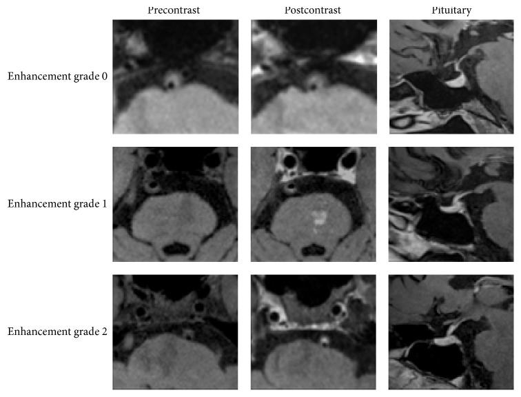
Fifty-seven patients (33 symptomatic patients and 24 asymptomatic patients) were recruited for 3.0 T HRMRI scan; all of them had >50% stenosis on the BA. The intraplaque hemorrhage (IPH), contrast-enhancement pattern, and distribution of BA plaques were compared between the symptomatic and asymptomatic groups. Factors potentially associated with posterior ischemic stroke were calculated by multivariate analyses.

RESULTS

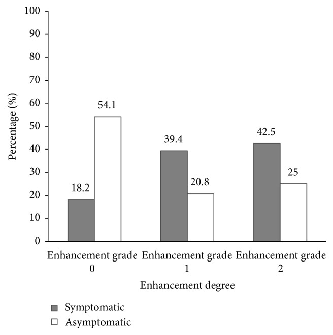
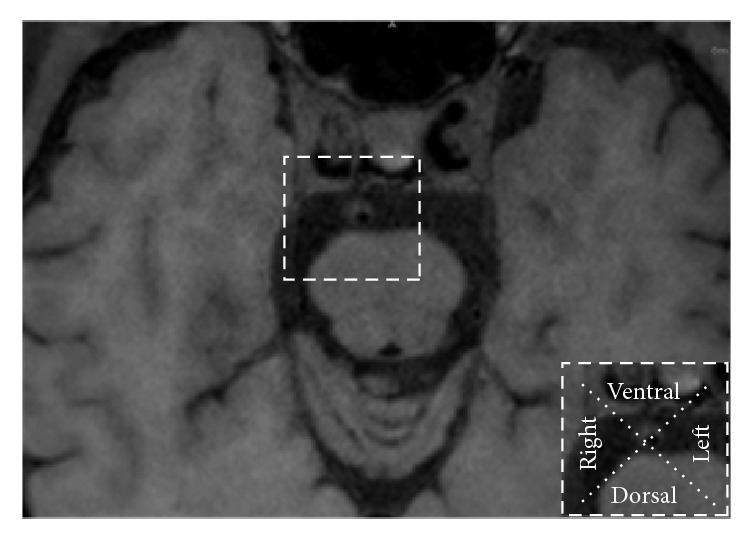
Enhancement of BA plaque was more frequently observed in symptomatic than in asymptomatic patients (27/33, 81.8% versus 11/24, 45.8%; < 0.01). In multivariate regression analysis, plaque enhancement (OR = 7.193; 95% CI: 1.880-27.517; = 0.004) and smoking (OR = 4.402; 95% CI: 2.218-15.909; = 0.024) were found to be independent risk factors of posterior ischemic events in patients with BA stenosis >50%. Plaques were mainly distributed at the ventral site (39.3%) or involved more than two arcs (21.2%) in the symptomatic group but were mainly distributed at left (33.3%) and right (25.0%) sites in the asymptomatic group.

摘要

目的

使用 3.0T 高分辨率磁共振成像(HRMRI)研究斑块的形态特征和分布模式与中重度基底动脉(BA)粥样硬化狭窄患者的临床相关性。

材料与方法

共招募 57 例患者(33 例有症状患者和 24 例无症状患者)进行 3.0T HRMRI 扫描;所有患者的 BA 狭窄率均>50%。比较有症状和无症状组患者的斑块内出血(IPH)、对比增强模式和 BA 斑块的分布情况。通过多变量分析计算与后部缺血性卒中相关的潜在因素。

结果

与无症状患者(24/24,45.8%)相比,有症状患者的 BA 斑块增强更为常见(27/33,81.8%)(<0.01)。多变量回归分析显示,斑块增强(OR=7.193;95%CI:1.880-27.517;=0.004)和吸烟(OR=4.402;95%CI:2.218-15.909;=0.024)是 BA 狭窄>50%患者发生后部缺血事件的独立危险因素。有症状组的斑块主要分布在腹侧部位(39.3%)或累及两个以上弧形(21.2%),而无症状组的斑块主要分布在左侧(33.3%)和右侧(25.0%)。

https://cdn.ncbi.nlm.nih.gov/pmc/blobs/db57/5623789/bb8014937250/BMRI2017-4281629.005.jpg
https://cdn.ncbi.nlm.nih.gov/pmc/blobs/db57/5623789/3676ce3587ce/BMRI2017-4281629.001.jpg
https://cdn.ncbi.nlm.nih.gov/pmc/blobs/db57/5623789/f827f3a56e4d/BMRI2017-4281629.002.jpg
https://cdn.ncbi.nlm.nih.gov/pmc/blobs/db57/5623789/1f5f7c5f7495/BMRI2017-4281629.003.jpg
https://cdn.ncbi.nlm.nih.gov/pmc/blobs/db57/5623789/de70613f1318/BMRI2017-4281629.004.jpg
https://cdn.ncbi.nlm.nih.gov/pmc/blobs/db57/5623789/bb8014937250/BMRI2017-4281629.005.jpg

相似文献

1
Incremental Value of Plaque Enhancement in Patients with Moderate or Severe Basilar Artery Stenosis: 3.0 T High-Resolution Magnetic Resonance Study.中重度基底动脉狭窄患者斑块强化的增量价值:3.0T 高分辨率磁共振研究。
Biomed Res Int. 2017;2017:4281629. doi: 10.1155/2017/4281629. Epub 2017 Sep 17.
2
Plaque distribution of low-grade basilar artery atherosclerosis and its clinical relevance.低级别基底动脉粥样硬化的斑块分布及其临床意义。
BMC Neurol. 2017 Jan 10;17(1):8. doi: 10.1186/s12883-016-0785-y.
3
Association of Intraplaque Hemorrhage and Acute Infarction in Patients With Basilar Artery Plaque.基底动脉斑块患者斑块内出血与急性梗死的相关性
Stroke. 2015 Oct;46(10):2768-72. doi: 10.1161/STROKEAHA.115.009412. Epub 2015 Aug 25.
4
Visualization of basilar artery atherosclerotic plaques by conventional T2-weighted magnetic resonance imaging: A case-control study.常规 T2 加权磁共振成像对基底动脉粥样硬化斑块的可视化:一项病例对照研究。
PLoS One. 2019 Feb 26;14(2):e0212570. doi: 10.1371/journal.pone.0212570. eCollection 2019.
5
The advantage of high-resolution MRI in evaluating basilar plaques: a comparison study with MRA.高分辨率 MRI 在评估基底节斑块中的优势:与 MRA 的对比研究。
Atherosclerosis. 2012 Oct;224(2):411-6. doi: 10.1016/j.atherosclerosis.2012.07.037. Epub 2012 Aug 13.
6
Clinical Significance of Intraplaque Hemorrhage in Low- and High-Grade Basilar Artery Stenosis on High-Resolution MRI.高分辨率 MRI 显示低级别和高级别基底动脉狭窄斑块内出血的临床意义。
AJNR Am J Neuroradiol. 2018 Jul;39(7):1286-1292. doi: 10.3174/ajnr.A5676. Epub 2018 May 24.
7
Morphologic characteristics of severe basilar artery atherosclerotic stenosis on 3D high-resolution MRI.三维高分辨率磁共振成像显示的重度基底动脉粥样硬化狭窄的形态学特征
BMC Neurol. 2018 Dec 15;18(1):206. doi: 10.1186/s12883-018-1214-1.
8
Association between basilar artery configuration and Vessel Wall features: a prospective high-resolution magnetic resonance imaging study.基底动脉形态与血管壁特征的相关性:一项前瞻性高分辨率磁共振成像研究。
BMC Med Imaging. 2019 Dec 26;19(1):99. doi: 10.1186/s12880-019-0388-3.
9
Characteristics of Symptomatic Basilar Artery Stenosis Using High-Resolution Magnetic Resonance Imaging in Ischemic Stroke Patients.症状性基底动脉狭窄的特征:基于缺血性脑卒中患者的高分辨率磁共振成像研究。
J Atheroscler Thromb. 2021 Oct 1;28(10):1063-1070. doi: 10.5551/jat.58214. Epub 2020 Nov 15.
10
Basilar artery atherosclerotic plaques distribution in symptomatic patients: a 3.0T high-resolution MRI study.症状性患者基底动脉粥样硬化斑块分布:3.0T 高分辨率 MRI 研究。
Eur J Radiol. 2013 Apr;82(4):e199-203. doi: 10.1016/j.ejrad.2012.10.031. Epub 2012 Dec 8.

引用本文的文献

1
Deep learning improves quality of intracranial vessel wall MRI for better characterization of potentially culprit plaques.深度学习提高颅内血管壁 MRI 质量,更好地对潜在罪犯斑块进行特征描述。
Sci Rep. 2024 Aug 16;14(1):18983. doi: 10.1038/s41598-024-69750-4.
2
Delayed Enhancement of Intracranial Atherosclerotic Plaque Can Better Differentiate Culprit Lesions: A Multiphase Contrast-Enhanced Vessel Wall MRI Study.颅内动脉粥样硬化斑块的延迟增强可以更好地区分罪犯病变:一项多期增强血管壁 MRI 研究。
AJNR Am J Neuroradiol. 2024 Mar 7;45(3):262-270. doi: 10.3174/ajnr.A8132.
3
Association between the fetal-type posterior cerebral artery and intracranial anterior and posterior circulating atherosclerotic plaques using multi-contrast magnetic resonance vessel wall imaging.

本文引用的文献

1
Plaque distribution of low-grade basilar artery atherosclerosis and its clinical relevance.低级别基底动脉粥样硬化的斑块分布及其临床意义。
BMC Neurol. 2017 Jan 10;17(1):8. doi: 10.1186/s12883-016-0785-y.
2
Magnetic Resonance Imaging of Plaque Morphology, Burden, and Distribution in Patients With Symptomatic Middle Cerebral Artery Stenosis.有症状大脑中动脉狭窄患者斑块形态、负荷及分布的磁共振成像
Stroke. 2016 Jul;47(7):1797-802. doi: 10.1161/STROKEAHA.116.013007. Epub 2016 Jun 14.
3
Vessel Wall Imaging of the Intracranial and Cervical Carotid Arteries.
利用多对比磁共振血管壁成像技术研究胎儿型大脑后动脉与颅内前后循环动脉粥样硬化斑块之间的关联。
Quant Imaging Med Surg. 2023 Dec 1;13(12):8383-8394. doi: 10.21037/qims-23-611. Epub 2023 Oct 27.
4
Association between basilar artery stenosis features, vertebral artery stenosis and perforator stroke after stenting.支架置入术后基底动脉狭窄特征、椎动脉狭窄与穿支卒中之间的关联。
Interv Neuroradiol. 2025 Apr;31(2):246-254. doi: 10.1177/15910199231164840. Epub 2023 Mar 29.
5
A Non-Linear Role of Hyperlipidemia on Progression of Intracranial Atherosclerotic Plaques and Acute Downstream Ischemic Events.高脂血症在颅内动脉粥样硬化斑块进展及急性下游缺血事件中的非线性作用
J Atheroscler Thromb. 2023 Oct 1;30(10):1448-1460. doi: 10.5551/jat.63971. Epub 2023 Jan 28.
6
High-risk features of basilar artery atherosclerotic plaque.基底动脉粥样硬化斑块的高危特征。
Front Neurol. 2022 Oct 28;13:1019036. doi: 10.3389/fneur.2022.1019036. eCollection 2022.
7
Characteristics of basilar artery atherosclerotic plaques in pontine infarctions: A high-resolution magnetic resonance imaging study.脑桥梗死中基底动脉粥样硬化斑块的特征:一项高分辨率磁共振成像研究。
Cereb Circ Cogn Behav. 2021 Feb 4;2:100005. doi: 10.1016/j.cccb.2021.100005. eCollection 2021.
8
A Predictive Model for the Risk of Posterior Circulation Stroke in Patients with Intracranial Atherosclerosis Based on High Resolution MRI.基于高分辨率磁共振成像的颅内动脉粥样硬化患者后循环卒中风险预测模型
Diagnostics (Basel). 2022 Mar 15;12(4):812. doi: 10.3390/diagnostics12040812.
9
Effect of Mid-Basilar Artery Angle and Plaque Characteristics on Pontine Infarction in Patients with Basilar Artery Plaque.基底动脉斑块患者中基底动脉中段夹角和斑块特征对脑桥梗死的影响。
J Atheroscler Thromb. 2023 Feb 1;30(2):182-191. doi: 10.5551/jat.63520. Epub 2022 Apr 13.
10
Baseline vessel wall magnetic resonance imaging characteristics associated with in-stent restenosis for intracranial atherosclerotic stenosis.颅内动脉粥样硬化狭窄支架内再狭窄与基线管壁磁共振成像特征的关系。
J Neurointerv Surg. 2023 Mar;15(3):288-291. doi: 10.1136/neurintsurg-2021-018473. Epub 2022 Mar 1.
颅内和颈内颈动脉血管壁成像。
J Stroke. 2015 Sep;17(3):238-55. doi: 10.5853/jos.2015.17.3.238. Epub 2015 Sep 30.
4
Association of Intraplaque Hemorrhage and Acute Infarction in Patients With Basilar Artery Plaque.基底动脉斑块患者斑块内出血与急性梗死的相关性
Stroke. 2015 Oct;46(10):2768-72. doi: 10.1161/STROKEAHA.115.009412. Epub 2015 Aug 25.
5
Characterization of Craniocervical Artery Dissection by Simultaneous MR Noncontrast Angiography and Intraplaque Hemorrhage Imaging at 3T.3T磁共振非对比血管造影和斑块内出血成像同步用于颅颈动脉夹层的特征分析
AJNR Am J Neuroradiol. 2015 Sep;36(9):1769-75. doi: 10.3174/ajnr.A4348. Epub 2015 Jun 4.
6
Basilar plaque on high-resolution MRI predicts progressive motor deficits after pontine infarction.高分辨率磁共振成像上的基底动脉斑块可预测脑桥梗死后的进行性运动功能缺损。
Atherosclerosis. 2015 May;240(1):278-83. doi: 10.1016/j.atherosclerosis.2015.03.029. Epub 2015 Mar 20.
7
Intracranial plaque enhancement in patients with cerebrovascular events on high-spatial-resolution MR images.高分辨率磁共振图像上脑血管事件患者颅内斑块增强。
Radiology. 2014 May;271(2):534-42. doi: 10.1148/radiol.13122812. Epub 2014 Jan 16.
8
Posterior circulation ischaemic stroke and transient ischaemic attack: diagnosis, investigation, and secondary prevention.后循环缺血性卒中和短暂性脑缺血发作:诊断、检查和二级预防。
Lancet Neurol. 2013 Oct;12(10):989-98. doi: 10.1016/S1474-4422(13)70211-4.
9
Basilar artery atherosclerotic plaques distribution in symptomatic patients: a 3.0T high-resolution MRI study.症状性患者基底动脉粥样硬化斑块分布:3.0T 高分辨率 MRI 研究。
Eur J Radiol. 2013 Apr;82(4):e199-203. doi: 10.1016/j.ejrad.2012.10.031. Epub 2012 Dec 8.
10
Clinical and histological significance of gadolinium enhancement in carotid atherosclerotic plaque.颈动脉粥样硬化斑块钆增强的临床和组织学意义。
Stroke. 2012 Nov;43(11):3023-8. doi: 10.1161/STROKEAHA.112.662692. Epub 2012 Aug 23.