• 文献检索
  • 文档翻译
  • 深度研究
  • 学术资讯
  • Suppr Zotero 插件Zotero 插件
  • 邀请有礼
  • 套餐&价格
  • 历史记录
应用&插件
Suppr Zotero 插件Zotero 插件浏览器插件Mac 客户端Windows 客户端微信小程序
定价
高级版会员购买积分包购买API积分包
服务
文献检索文档翻译深度研究API 文档MCP 服务
关于我们
关于 Suppr公司介绍联系我们用户协议隐私条款
关注我们

Suppr 超能文献

核心技术专利:CN118964589B侵权必究
粤ICP备2023148730 号-1Suppr @ 2026

文献检索

告别复杂PubMed语法,用中文像聊天一样搜索,搜遍4000万医学文献。AI智能推荐,让科研检索更轻松。

立即免费搜索

文件翻译

保留排版,准确专业,支持PDF/Word/PPT等文件格式,支持 12+语言互译。

免费翻译文档

深度研究

AI帮你快速写综述,25分钟生成高质量综述,智能提取关键信息,辅助科研写作。

立即免费体验

原发性食管癌中,通过诊断性PET/CT与计划CT的可变形配准和刚性配准所定义的靶区体积的比较评估。

Comparative evaluation of target volumes defined by deformable and rigid registration of diagnostic PET/CT to planning CT in primary esophageal cancer.

作者信息

Guo Yanluan, Li Jianbin, Zhang Peng, Shao Qian, Xu Min, Li Yankang

机构信息

Department of Radiation Oncology, Shandong Cancer Hospital and Institute, Jinan, Shandong, China.

出版信息

Medicine (Baltimore). 2017 Jan;96(1):e5528. doi: 10.1097/MD.0000000000005528.

DOI:10.1097/MD.0000000000005528
PMID:28072693
原文链接:https://pmc.ncbi.nlm.nih.gov/articles/PMC5228653/
Abstract

BACKGROUND

To evaluate the geometrical differences of target volumes propagated by deformable image registration (DIR) and rigid image registration (RIR) to assist target volume delineation between diagnostic Positron emission tomography/computed tomography (PET/CT) and planning CT for primary esophageal cancer (EC).

METHODS

Twenty-five patients with EC sequentially underwent a diagnostic F-fluorodeoxyglucose (F-FDG) PET/CT scan and planning CT simulation. Only 19 patients with maximum standardized uptake value (SUVmax) ≥ 2.0 of the primary volume were available. Gross tumor volumes (GTVs) were delineated using CT and PET display settings. The PET/CT images were then registered with planning CT using MIM software. Subsequently, the PET and CT contours were propagated by RIR and DIR to planning CT. The properties of these volumes were compared.

RESULTS

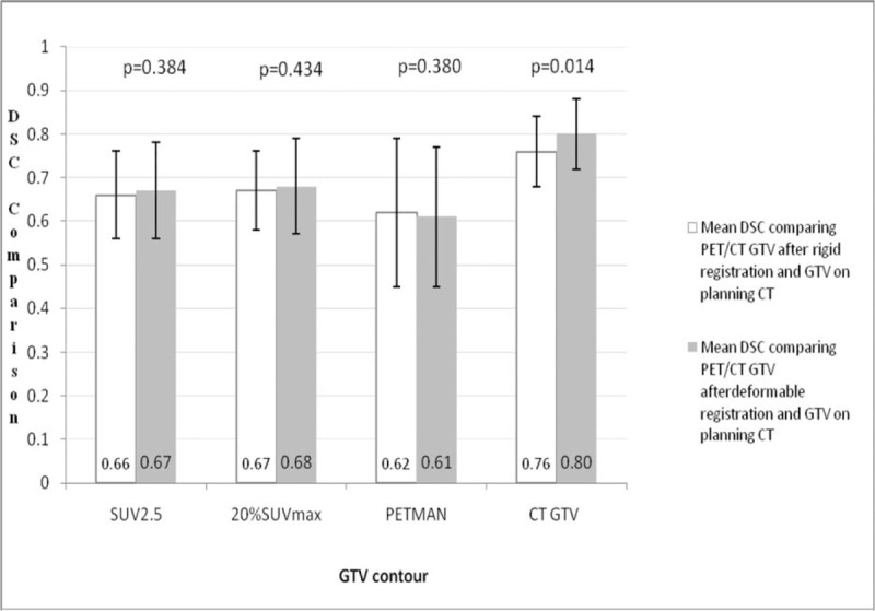
When GTVCT delineated on CT of PET/CT after both RIR and DIR was compared with GTV contoured on planning CT, significant improvements using DIR were observed in the volume, displacements of the center of mass (COM) in the 3-dimensional (3D) direction, and Dice similarity coefficient (DSC) (P = 0.003; 0.006; 0.014). Although similar improvements were not observed for the same comparison using DIR for propagated PET contours from diagnostic PET/CT to planning CT (P > 0.05), for DSC and displacements of COM in the 3D direction of PET contours, the DIR resulted in the improved volume of a large percentage of patients (73.7%; 68.45%; 63.2%) compared with RIR. For diagnostic CT-based contours or PET contours at SUV2.5 propagated by DIR with planning CT, the DSC and displacements of COM in 3D directions in the distal segment were significantly improved compared to the upper and middle segments (P > 0.05).

CONCLUSION

We observed a trend that deformable registration might improve the overlap for gross target volumes from diagnostic PET/CT to planning CT. The distal EC might benefit more from DIR.

摘要

背景

评估通过可变形图像配准(DIR)和刚性图像配准(RIR)传播的靶区体积的几何差异,以辅助原发性食管癌(EC)诊断性正电子发射断层扫描/计算机断层扫描(PET/CT)与计划CT之间的靶区体积勾画。

方法

25例EC患者依次接受诊断性F-氟脱氧葡萄糖(F-FDG)PET/CT扫描和计划CT模拟。仅19例原发灶最大标准化摄取值(SUVmax)≥2.0的患者可用。使用CT和PET显示设置勾画大体肿瘤体积(GTV)。然后使用MIM软件将PET/CT图像与计划CT配准。随后,通过RIR和DIR将PET和CT轮廓传播到计划CT。比较这些体积的属性。

结果

当将RIR和DIR后PET/CT的CT上勾画的GTVCT与计划CT上勾画的GTV进行比较时,在体积、三维(3D)方向上质心(COM)的位移和骰子相似系数(DSC)方面,使用DIR观察到显著改善(P = 0.003;0.006;0.014)。虽然从诊断性PET/CT到计划CT传播的PET轮廓使用DIR进行相同比较时未观察到类似改善(P>0.05),但对于PET轮廓的DSC和3D方向上COM的位移,与RIR相比,DIR使很大比例的患者体积得到改善(73.7%;68.45%;63.2%)。对于基于诊断CT的轮廓或通过DIR与计划CT传播的SUV2.5处的PET轮廓,与上段和中段相比,远端节段的DSC和3D方向上COM的位移显著改善(P>0.05)。

结论

我们观察到一种趋势,即可变形配准可能会改善从诊断性PET/CT到计划CT的大体靶区体积的重叠。远端EC可能从DIR中获益更多。

https://cdn.ncbi.nlm.nih.gov/pmc/blobs/1f24/5228653/e141336ac872/medi-96-e5528-g025.jpg

相似文献

1
Comparative evaluation of target volumes defined by deformable and rigid registration of diagnostic PET/CT to planning CT in primary esophageal cancer.原发性食管癌中,通过诊断性PET/CT与计划CT的可变形配准和刚性配准所定义的靶区体积的比较评估。
Medicine (Baltimore). 2017 Jan;96(1):e5528. doi: 10.1097/MD.0000000000005528.
2
Superiority of Deformable Image Co-registration in the Integration of Diagnostic Positron Emission Tomography-Computed Tomography to the Radiotherapy Treatment Planning Pathway for Oesophageal Carcinoma.在食管癌放射治疗计划流程中,可变形图像配准在诊断性正电子发射断层扫描-计算机断层扫描整合中的优势
Clin Oncol (R Coll Radiol). 2016 Oct;28(10):655-62. doi: 10.1016/j.clon.2016.05.009. Epub 2016 Jun 3.
3
Comparison of rigid and deformable image registration for nasopharyngeal carcinoma radiotherapy planning with diagnostic position PET/CT.比较诊断体位 PET/CT 引导下鼻咽癌放疗计划中刚性和弹性图像配准。
Jpn J Radiol. 2020 Mar;38(3):256-264. doi: 10.1007/s11604-019-00911-6. Epub 2019 Dec 13.
4
[F18] FDG-PET/CT for manual or semiautomated GTV delineation of the primary tumor for radiation therapy planning in patients with esophageal cancer: is it useful?[F18] 氟代脱氧葡萄糖正电子发射断层扫描/计算机断层扫描用于手动或半自动勾画食管癌放射治疗计划中肿瘤原发灶的大体肿瘤靶区:是否有用?
Strahlenther Onkol. 2021 Sep;197(9):780-790. doi: 10.1007/s00066-020-01701-0. Epub 2020 Oct 26.
5
Conventional 3D staging PET/CT in CT simulation for lung cancer: impact of rigid and deformable target volume alignments for radiotherapy treatment planning.肺癌 CT 模拟中常规 3D 分期 PET/CT:刚性和弹性靶区配准对放疗计划的影响。
Br J Radiol. 2011 Oct;84(1006):919-29. doi: 10.1259/bjr/29163167. Epub 2011 Jan 11.
6
Geometrical differences in target volumes based on 18F-fluorodeoxyglucose positron emission tomography/computed tomography and four-dimensional computed tomography maximum intensity projection images of primary thoracic esophageal cancer.基于18F-氟脱氧葡萄糖正电子发射断层扫描/计算机断层扫描及原发性胸段食管癌的四维计算机断层扫描最大密度投影图像的靶区几何差异。
Dis Esophagus. 2014 Nov-Dec;27(8):744-50. doi: 10.1111/dote.12247. Epub 2014 Jun 11.
7
Tumour delineation in oesophageal cancer - A prospective study of delineation in PET and CT with and without endoscopically placed clip markers.食管癌肿瘤勾画 - 内镜放置夹标记 PET 和 CT 勾画的前瞻性研究。
Radiother Oncol. 2015 Aug;116(2):269-75. doi: 10.1016/j.radonc.2015.07.007. Epub 2015 Sep 10.
8
Evaluation of a commercial DIR platform for contour propagation in prostate cancer patients treated with IMRT/VMAT.评价一个商业的 DIR 平台在接受调强放疗/VMAT 治疗的前列腺癌患者中的靶区勾画。
J Appl Clin Med Phys. 2020 Feb;21(2):14-25. doi: 10.1002/acm2.12787.
9
Comparison of rigid and deformable registration through the respiratory phases of four-dimensional computed tomography image data sets for radiotherapy after breast-conserving surgery.通过保乳手术后放疗的四维计算机断层扫描图像数据集的呼吸阶段比较刚性配准和可变形配准。
Medicine (Baltimore). 2017 Dec;96(50):e9143. doi: 10.1097/MD.0000000000009143.
10
Comparison of gross target volumes based on four-dimensional CT, positron emission tomography-computed tomography, and magnetic resonance imaging in thoracic esophageal cancer.基于 4DCT、PET-CT 和 MRI 的胸段食管癌大体肿瘤靶区体积比较。
Cancer Med. 2020 Aug;9(15):5353-5361. doi: 10.1002/cam4.3072. Epub 2020 Jun 8.

引用本文的文献

1
Gross Tumor Volume Definition and Comparative Assessment for Esophageal Squamous Cell Carcinoma From 3D F-FDG PET/CT by Deep Learning-Based Method.基于深度学习方法的3D F-FDG PET/CT对食管鳞状细胞癌的肿瘤总体积定义及比较评估
Front Oncol. 2022 Mar 17;12:799207. doi: 10.3389/fonc.2022.799207. eCollection 2022.
2
Comparison of the Gross Target Volumes Based on Diagnostic PET/CT for Primary Esophageal Cancer.基于诊断性PET/CT的原发性食管癌大体靶体积比较。
Front Oncol. 2021 Feb 25;11:550100. doi: 10.3389/fonc.2021.550100. eCollection 2021.
3
Role of clip markers placed by endoscopic ultrasonography in contouring gross tumor volume for thoracic esophageal squamous cell carcinoma: one prospective study.

本文引用的文献

1
Intrafraction variability and deformation quantification in the breast.乳腺内分次照射的可变性与形变量化
Int J Radiat Oncol Biol Phys. 2015 Mar 1;91(3):604-11. doi: 10.1016/j.ijrobp.2014.11.003. Epub 2015 Jan 30.
2
PET/CT imaging-guided dose painting in radiation therapy.正电子发射断层扫描/计算机断层扫描成像引导下的放射治疗剂量描绘。
Cancer Lett. 2014 Dec 28;355(2):169-75. doi: 10.1016/j.canlet.2014.07.042. Epub 2014 Sep 10.
3
Deformable versus rigid registration of PET/CT images for radiation treatment planning of head and neck and lung cancer patients: a retrospective dosimetric comparison.
内镜超声放置的夹子标记物在勾画胸段食管鳞状细胞癌大体肿瘤体积中的作用:一项前瞻性研究
Ann Transl Med. 2020 Sep;8(18):1144. doi: 10.21037/atm-20-4030.
4
Comparison of different images in gross target volume delineating on VX2 nasopharyngeal transplantation tumor models.VX2鼻咽癌移植瘤模型大体靶区勾画中不同图像的比较
J Cancer. 2020 Jan 1;11(5):1104-1114. doi: 10.7150/jca.36076. eCollection 2020.
5
Normal liver tissue change after proton beam therapy.质子束治疗后正常肝组织的变化。
Jpn J Radiol. 2018 Sep;36(9):559-565. doi: 10.1007/s11604-018-0757-9. Epub 2018 Jul 6.
用于头颈癌和肺癌患者放射治疗计划的PET/CT图像的可变形配准与刚性配准:一项回顾性剂量学比较
Radiat Oncol. 2014 Feb 10;9:50. doi: 10.1186/1748-717X-9-50.
4
Deformable registration of preoperative PET/CT with postoperative radiation therapy planning CT in head and neck cancer.头颈部癌症中术前 PET/CT 与术后放射治疗计划 CT 的可变形配准。
Radiographics. 2012 Sep-Oct;32(5):1329-41. doi: 10.1148/rg.325125008.
5
The clinical application of 4D 18F-FDG PET/CT on gross tumor volume delineation for radiotherapy planning in esophageal squamous cell cancer.4D18F-FDG PET/CT 在食管鳞癌放疗计划中大体肿瘤体积勾画的临床应用。
J Radiat Res. 2012 Jul;53(4):594-600. doi: 10.1093/jrr/rrs009. Epub 2012 Jun 5.
6
Comparison of the planning target volume based on three-dimensional CT and four-dimensional CT images of non-small-cell lung cancer.非小细胞肺癌三维 CT 和四维 CT 图像的计划靶区比较。
Radiother Oncol. 2011 May;99(2):176-80. doi: 10.1016/j.radonc.2011.03.015. Epub 2011 May 4.
7
The evaluation of a deformable image registration segmentation technique for semi-automating internal target volume (ITV) production from 4DCT images of lung stereotactic body radiotherapy (SBRT) patients.一种用于从肺部立体定向体部放疗(SBRT)患者的 4DCT 图像半自动生成内部靶区(ITV)的可变形图像配准分割技术的评估。
Radiother Oncol. 2011 Feb;98(2):277-83. doi: 10.1016/j.radonc.2010.12.007. Epub 2011 Jan 21.
8
Conventional 3D staging PET/CT in CT simulation for lung cancer: impact of rigid and deformable target volume alignments for radiotherapy treatment planning.肺癌 CT 模拟中常规 3D 分期 PET/CT:刚性和弹性靶区配准对放疗计划的影响。
Br J Radiol. 2011 Oct;84(1006):919-29. doi: 10.1259/bjr/29163167. Epub 2011 Jan 11.
9
Computed tomography overestimation of esophageal tumor length: Implications for radiotherapy planning.计算机断层扫描高估食管肿瘤长度:对放射治疗计划的影响。
World J Gastrointest Oncol. 2010 Apr 15;2(4):197-204. doi: 10.4251/wjgo.v2.i4.197.
10
PET-guided delineation of radiation therapy treatment volumes: a survey of image segmentation techniques.正电子发射断层扫描(PET)引导的放射治疗靶区勾画:图像分割技术调查。
Eur J Nucl Med Mol Imaging. 2010 Nov;37(11):2165-87. doi: 10.1007/s00259-010-1423-3. Epub 2010 Mar 25.