Glausier Jill R, Roberts Rosalinda C, Lewis David A
Department of Psychiatry, University of Pittsburgh School of Medicine, Pittsburgh, Pennsylvania.
Department of Psychiatry and Behavioral Neurobiology, University of Alabama, Birmingham, Alabama.
J Comp Neurol. 2017 Jun 15;525(9):2075-2089. doi: 10.1002/cne.24171. Epub 2017 Mar 26.
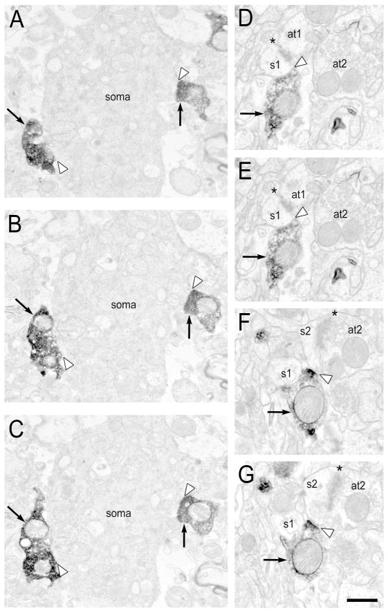
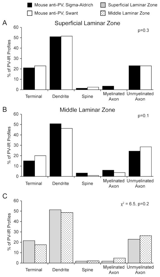
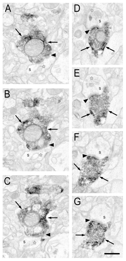
Coordinated activity of neural circuitry in the primate dorsolateral prefrontal cortex (DLPFC) supports a range of cognitive functions. Altered DLPFC activation is implicated in a number of human psychiatric and neurological illnesses. Proper DLPFC activity is, in part, maintained by two populations of neurons containing the calcium-binding protein parvalbumin (PV): local inhibitory interneurons that form Type II synapses, and long-range glutamatergic inputs from the thalamus that form Type I synapses. Understanding the contributions of each PV neuronal population to human DLPFC function requires a detailed examination of their anatomical properties. Consequently, we performed an electron microscopic analysis of (1) the distribution of PV immunoreactivity within the neuropil, (2) the properties of dendritic shafts of PV-IR interneurons, (3) Type II PV-IR synapses from PV interneurons, and (4) Type I PV-IR synapses from long-range projections, within the superficial and middle laminar zones of the human DLPFC. In both laminar zones, Type II PV-IR synapses from interneurons comprised ∼60% of all PV-IR synapses, and Type I PV-IR synapses from putative thalamocortical terminals comprised the remaining ∼40% of PV-IR synapses. Thus, the present study suggests that innervation from PV-containing thalamic nuclei extends across superficial and middle layers of the human DLPFC. These findings contrast with previous ultrastructural studies in monkey DLPFC where Type I PV-IR synapses were not identified in the superficial laminar zone. The presumptive added modulation of DLPFC circuitry by the thalamus in human may contribute to species-specific, higher-order functions.
灵长类动物背外侧前额叶皮层(DLPFC)中神经回路的协调活动支持一系列认知功能。DLPFC激活的改变与许多人类精神和神经疾病有关。适当的DLPFC活动部分由两类含有钙结合蛋白小白蛋白(PV)的神经元维持:形成II型突触的局部抑制性中间神经元,以及来自丘脑形成I型突触的长距离谷氨酸能输入。了解每个PV神经元群体对人类DLPFC功能的贡献需要详细检查它们的解剖学特性。因此,我们对人类DLPFC浅层和中层区域内的(1)神经毡内PV免疫反应性的分布、(2)PV免疫反应性中间神经元树突干的特性、(3)PV中间神经元的II型PV免疫反应性突触和(4)长距离投射的I型PV免疫反应性突触进行了电子显微镜分析。在这两个层区,中间神经元的II型PV免疫反应性突触约占所有PV免疫反应性突触的60%,推测丘脑皮质终末的I型PV免疫反应性突触约占PV免疫反应性突触的其余40%。因此,本研究表明,来自含PV丘脑核的神经支配延伸至人类DLPFC的浅层和中层。这些发现与之前对猴DLPFC的超微结构研究形成对比,在猴DLPFC的浅层区域未发现I型PV免疫反应性突触。人类丘脑对DLPFC神经回路可能的额外调节可能有助于物种特异性的高阶功能。