Lloyd Adam, Navarrete Geraldine, Marchesan Melissa Andreia, Clement David
- University of Tennessee Health Science Center, College of Dentistry, Department of Endodontics, Memphis, TN, USA.
- The University of Oklahoma, College of Dentistry, Oklahoma City, OK, USA.
J Appl Oral Sci. 2016 Nov-Dec;24(6):543-548. doi: 10.1590/1678-775720160234.
This study compared the effectiveness of Er:YAG laser-activated irrigation (PIPS), passive ultrasonic irrigation (PUI) with EndoUltra and standard needle irrigation (SNI) in the removal of calcium hydroxide [Ca(OH)2] from the mesial roots of Weine Type II mandibular molars.
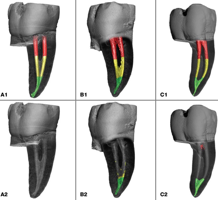
Thirty mandibular molars were screened by µCT for the presence of mesial roots with complex intra-canal anatomy and a common apical foramen. The teeth were enlarged to a standardized 25/.06 preparation and filled with Ca(OH)2 paste. Specimens were divided into three groups (n=10) according to the technique used for Ca(OH)2 removal: PIPS, at 15 Hz and 20 mJ using a 9 mm long, 600 µm diameter tip; PUI using a 15/.02 tip; and SNI (30 Ga. side-vented needle). Equal volumes of 8.25% NaOCl and 17% EDTA were used in all groups. µCT was used to measure the initial amount of Ca(OH)2 present and to assess the residual volume of Ca(OH)2 following each irrigation protocol. Data were analyzed using Tukey HSD and Kruskal-Wallis tests (α=5%).
The mean volume of Ca(OH)2 before removal was significantly higher in the coronal third than in the middle and apical third (p<0.001). Ca(OH)2 was similarly removed from the coronal and middle thirds with the three methods used (p>0.05). PIPS (median 0%; IQR: 0-0) showed significant higher Ca(OH)2 removal in the apical third than PUI (median 100%, IQR: 85-100) and SNI (median 47%; IQR: 16-72) (p<0.001).
PIPS laser-activation was more effective for the removal of Ca(OH)2 from mesial roots of mandibular molars with Weine Type II canal configurations than PUI with EndoUltra and SNI.
本研究比较了铒钇铝石榴石(Er:YAG)激光激活冲洗(PIPS)、使用EndoUltra的被动超声冲洗(PUI)和标准针管冲洗(SNI)从Weine II型下颌磨牙近中根去除氢氧化钙[Ca(OH)₂]的效果。
通过显微CT筛选出30颗下颌磨牙,其近中根具有复杂的根管解剖结构且根尖孔相通。将牙齿扩大至标准化的25/.06预备规格,并用Ca(OH)₂糊剂充填。根据去除Ca(OH)₂所使用的技术,将标本分为三组(n = 10):PIPS组,使用9毫米长、600微米直径的尖端,频率为15赫兹,能量为20毫焦;PUI组,使用15/.02的尖端;SNI组(30号侧孔针)。所有组均使用等量的8.25%次氯酸钠和17%乙二胺四乙酸。使用显微CT测量Ca(OH)₂的初始含量,并评估每种冲洗方案后Ca(OH)₂的残留量。使用Tukey HSD检验和Kruskal-Wallis检验对数据进行分析(α = 5%)。
去除前,冠方三分之一处Ca(OH)₂的平均体积显著高于中部和根尖三分之一处(p < 0.001)。使用的三种方法从冠方和中部三分之一处去除Ca(OH)₂的情况相似(p > 0.05)。PIPS组(中位数0%;四分位间距:0 - 0)在根尖三分之一处去除Ca(OH)₂的效果显著高于PUI组(中位数100%,四分位间距:85 - 100)和SNI组(中位数47%;四分位间距:16 - 72)(p < 0.001)。
对于具有Weine II型根管形态的下颌磨牙近中根而言,PIPS激光激活在去除Ca(OH)₂方面比使用EndoUltra的PUI和SNI更有效。