Bajaj Sahil, Housley Stephen N, Wu David, Dhamala Mukesh, James G A, Butler Andrew J
Department of Physics and Astronomy, Georgia State University, AtlantaGA, USA; Department of Psychiatry, College of Medicine, University of Arizona, TucsonAZ, USA.
Byrdine F. Lewis School of Nursing and Health Professions, Georgia State University, Atlanta GA, USA.
Front Hum Neurosci. 2016 Dec 27;10:650. doi: 10.3389/fnhum.2016.00650. eCollection 2016.
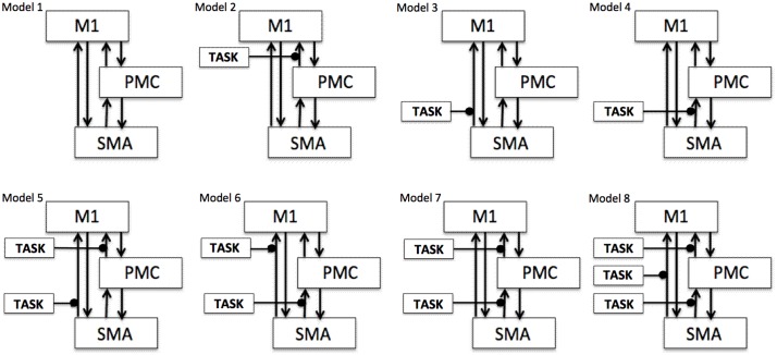
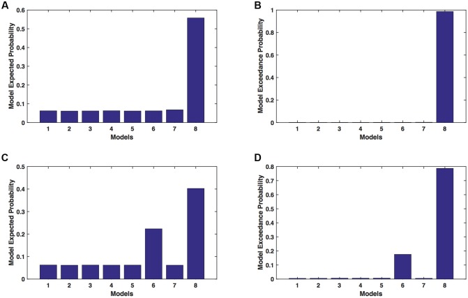
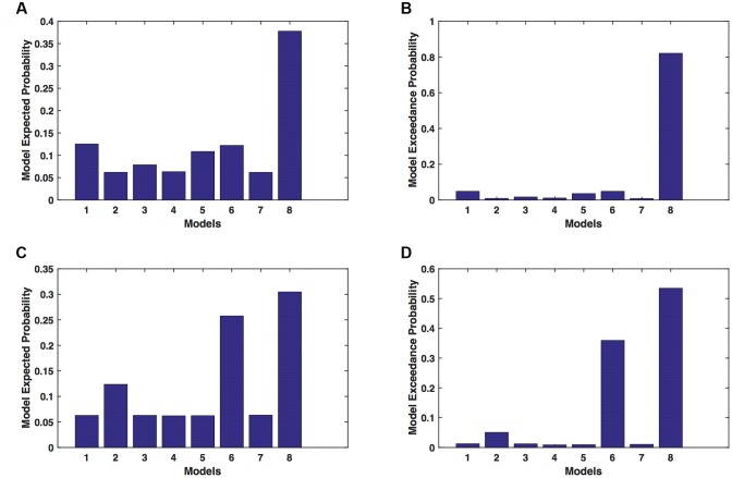
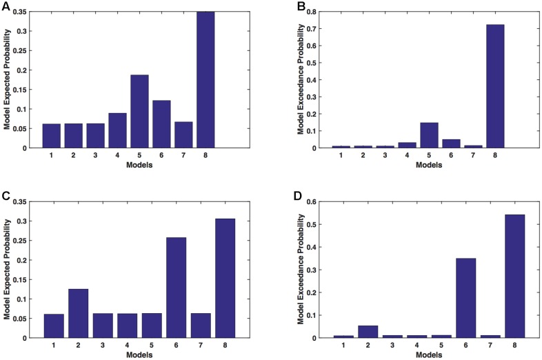
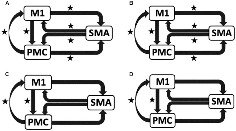
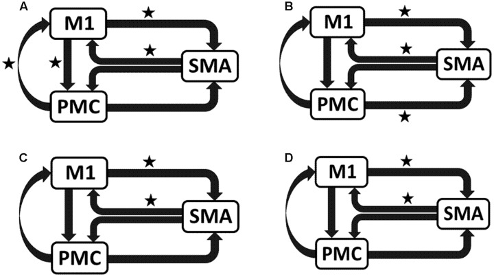
Balance of motor network activity between the two brain hemispheres after stroke is crucial for functional recovery. Several studies have extensively studied the role of the affected brain hemisphere to better understand changes in motor network activity following stroke. Very few studies have examined the role of the unaffected brain hemisphere and confirmed the test-retest reliability of connectivity measures on unaffected hemisphere. We recorded blood oxygenation level dependent functional magnetic resonance imaging (fMRI) signals from nine stroke survivors with hemiparesis of the left or right hand. Participants performed a motor execution task with affected hand, unaffected hand, and both hands simultaneously. Participants returned for a repeat fMRI scan 1 week later. Using dynamic causal modeling (DCM), we evaluated effective connectivity among three motor areas: the primary motor area (M1), the premotor cortex (PMC) and the supplementary motor area for the affected and unaffected hemispheres separately. Five participants' manual motor ability was assessed by Fugl-Meyer Motor Assessment scores and root-mean square error of participants' tracking ability during a robot-assisted game. We found (i) that the task performance with the affected hand resulted in strengthening of the connectivity pattern for unaffected hemisphere, (ii) an identical network of the unaffected hemisphere when participants performed the task with their unaffected hand, and (iii) the pattern of directional connectivity observed in the affected hemisphere was identical for tasks using the affected hand only or both hands. Furthermore, paired -test comparison found no significant differences in connectivity strength for any path when compared with one-week follow-up. Brain-behavior linear correlation analysis showed that the connectivity patterns in the unaffected hemisphere more accurately reflected the behavioral conditions than the connectivity patterns in the affected hemisphere. Above findings enrich our knowledge of unaffected brain hemisphere following stroke, which further strengthens our neurobiological understanding of stroke-affected brain and can help to effectively identify and apply stroke-treatments.
中风后两个脑半球之间运动网络活动的平衡对于功能恢复至关重要。多项研究广泛探讨了受影响脑半球的作用,以更好地理解中风后运动网络活动的变化。很少有研究考察未受影响脑半球的作用,也未证实未受影响半球连接性测量的重测信度。我们记录了9名左手或右手偏瘫的中风幸存者的血氧水平依赖性功能磁共振成像(fMRI)信号。参与者用患手、健手以及双手同时执行一项运动执行任务。1周后,参与者返回进行重复的fMRI扫描。我们使用动态因果模型(DCM)分别评估了受影响和未受影响半球的三个运动区域之间的有效连接性:初级运动区(M1)、运动前皮质(PMC)和辅助运动区。通过Fugl-Meyer运动评估分数和参与者在机器人辅助游戏中的跟踪能力的均方根误差评估了5名参与者的手动运动能力。我们发现:(i)患手的任务表现导致未受影响半球的连接模式增强;(ii)当参与者用健手执行任务时,未受影响半球的网络相同;(iii)仅使用患手或双手执行任务时,在受影响半球观察到的定向连接模式相同。此外,配对检验比较发现,与一周后的随访相比,任何路径的连接强度均无显著差异。脑-行为线性相关分析表明,未受影响半球的连接模式比受影响半球的连接模式更准确地反映了行为状况。上述发现丰富了我们对中风后未受影响脑半球的认识,进一步加强了我们对中风影响大脑的神经生物学理解,并有助于有效地识别和应用中风治疗方法。