Green Nancy S, Bhatia Monica, Griffith Erica Y, Qureshi Mahvish, Briamonte Courtney, Savone Mirko, Sands Stephen, Lee Margaret T, Lignelli Angela, Brickman Adam M
Department of Pediatrics, Columbia University Medical Center, New York, New York.
Department of Pediatrics, Columbia University Medical Center, New York, New York.
Biol Blood Marrow Transplant. 2017 Apr;23(4):670-676. doi: 10.1016/j.bbmt.2017.01.007. Epub 2017 Jan 9.
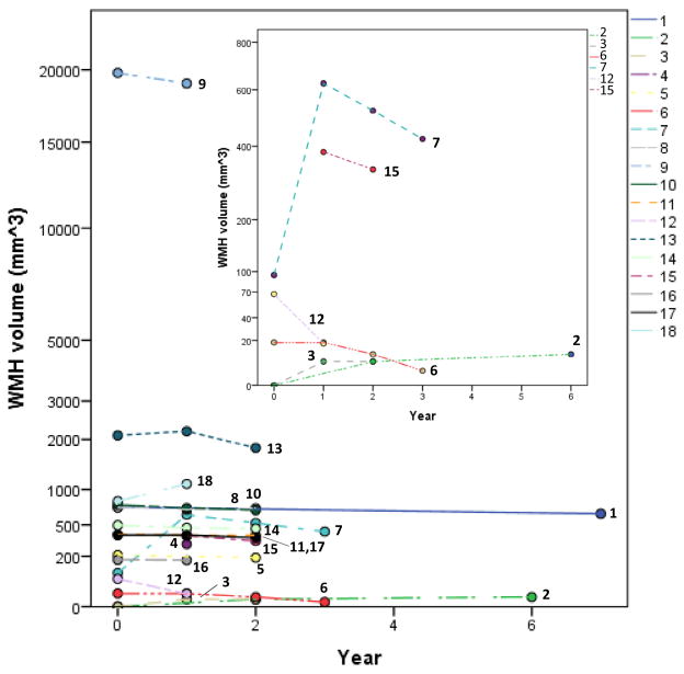
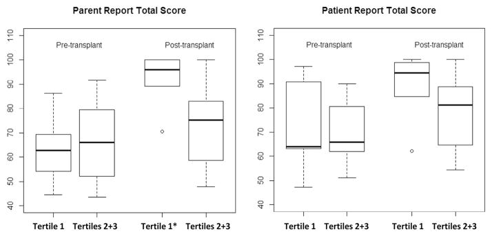
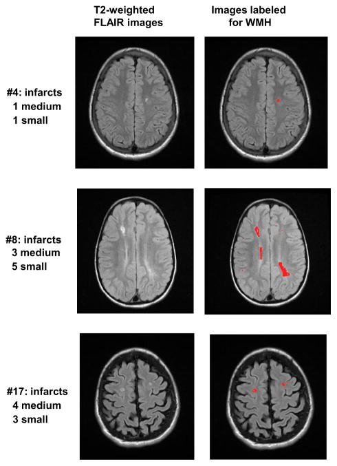
Progressive neurovasculopathy in children with sickle cell disease (SCD) results in decreased cognitive function and quality of life (QoL). Hematopoietic cell transplantation (HCT) is believed to halt progression of neurovasculopathy. Quantitative analysis of T2-weighted fluid attenuated inversion recovery (FLAIR) magnetic resonance imaging (MRI) for white matter hyperintensity (WMH) burden provides a meaningful estimate of small vessel cerebrovascular disease. We asked if quantitative analysis of WMH could complement standardized clinical assessment of MRI/magnetic resonance angiography (MRA) for assessing SCD central nervous system vasculopathy before and after HCT. Retrospective longitudinal clinical examination of scheduled annual MRI/MRA and quantitative analysis of WMH were performed before and 1 to 7 years after HCT at scheduled annual intervals, along with QoL measurements, in children who had engrafted after HCT. Of 18 patients alive and persistently engrafted (median age, 9.1 years), pretransplantation MRI demonstrated that 9 and 5 had sickle-related stroke and/or small infarcts, respectively. Patients were divided into WMH severity tertiles based on pretransplantation WMH volumes. MRI and WMH were assessed 1 to 7 years after HCT. MRI/MRA and WMH volume were stable or slightly better in 17 of 18 patients. By parent- and self-report, post-HCT QoL improved for children in the lowest WMH tertile significantly more than in the other groups. Based on this single-institution retrospective sample, we report that WMH appears to quantitatively support MRI-based findings that HCT stabilizes long-term small and large vessel cerebrovascular changes and is associated with the degree of improved QoL. While confirmation in larger prospective studies and evaluation by neurocognitive testing are needed, these findings suggest that WMH is a useful biomarker of neurovasculopathy after transplantation for SCD.
镰状细胞病(SCD)患儿的进行性神经血管病变会导致认知功能和生活质量(QoL)下降。造血细胞移植(HCT)被认为可以阻止神经血管病变的进展。对白质高信号(WMH)负荷进行T2加权液体衰减反转恢复(FLAIR)磁共振成像(MRI)定量分析,可为小血管脑血管疾病提供有意义的评估。我们探讨了WMH定量分析是否能补充MRI/磁共振血管造影(MRA)的标准化临床评估,以评估HCT前后SCD中枢神经系统血管病变。对接受HCT后植入成功的儿童,在预定的年度间隔时间进行回顾性纵向临床检查,包括每年的MRI/MRA检查以及WMH定量分析,并在HCT前和HCT后1至7年进行生活质量测量。在18名存活且持续植入成功的患者(中位年龄9.1岁)中,移植前MRI显示分别有9例和5例患有镰状细胞相关中风和/或小梗死。根据移植前WMH体积将患者分为WMH严重程度三分位数组。在HCT后1至7年评估MRI和WMH。18名患者中有17名的MRI/MRA和WMH体积稳定或略有改善。通过家长和自我报告,WMH三分位数最低组的儿童HCT后的生活质量改善明显超过其他组。基于这个单机构回顾性样本,我们报告WMH似乎在定量上支持基于MRI的研究结果,即HCT可稳定长期的小血管和大血管脑血管变化,并与生活质量改善程度相关。虽然需要在更大规模的前瞻性研究中进行验证并通过神经认知测试进行评估,但这些发现表明WMH是SCD移植后神经血管病变的有用生物标志物。