van der Bijl Pieter, Herbst Philip, Doubell Anton F
Division of Cardiology, Department of Medicine, Faculty of Medicine and Health Sciences, Stellenbosch University and Tygerberg Academic Hospital, Parow, South Africa.
J Cardiovasc Ultrasound. 2016 Dec;24(4):317-323. doi: 10.4250/jcu.2016.24.4.317. Epub 2016 Dec 28.
Effusive-constrictive pericarditis (ECP) is traditionally diagnosed by using the expensive and invasive technique of direct pressure measurements in the pericardial space and the right atrium. The aim of this study was to assess the diagnostic role of echocardiography in tuberculous ECP.
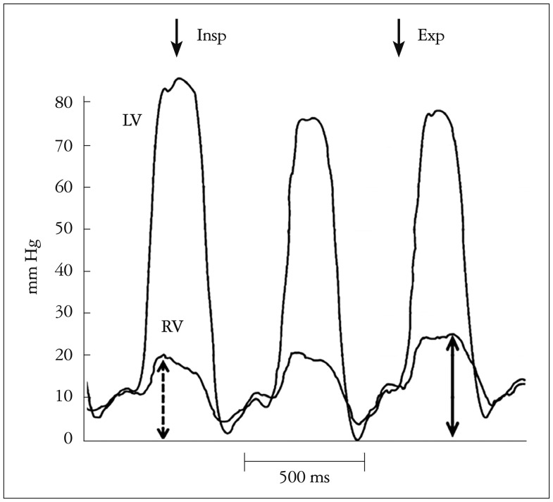
Intrapericardial and right atrial pressures were measured pre- and post-pericardiocentesis, and right ventricular and left ventricular pressures were measured post-pericardiocentesis in patients with tuberculous pericardial effusions. Echocardiography was performed post-pericardiocentesis. Traditional, pressure-based diagnostic criteria were compared with post-pericardiocentesis systolic discordance and echocardiographic evidence of constriction.
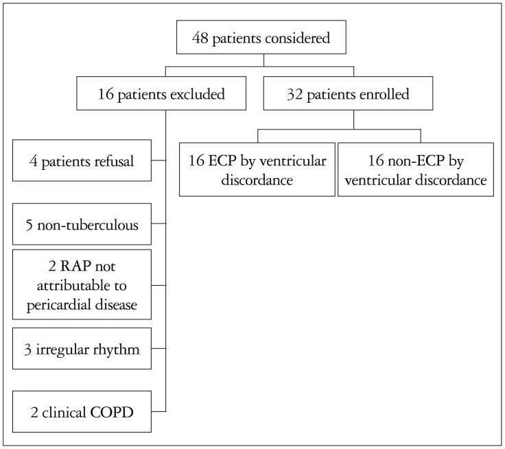
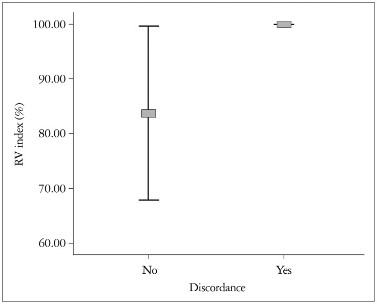
Thirty-two patients with tuberculous pericardial disease were included. Sixteen had ventricular discordance (invasively measured), 16 had ECP as measured by intrapericardial and right atrial invasive pressure measurements and 17 had ECP determined echocardiographically. The sensitivity and specificity of pressure-guided measurements (compared with discordance) for the diagnosis of ECP were both 56%. The positive and negative predictive values were both 56%. The sensitivity of echocardiography (compared with discordance) for the diagnosis of ECP was 81% and the specificity 75%, while the positive and the negative predictive values were 76% and 80%, respectively.
Echocardiography shows a better diagnostic performance than invasive, pressure-based measurements for the diagnosis of ECP when both these techniques are compared with the gold standard of invasively measured systolic discordance.
传统上,渗出性缩窄性心包炎(ECP)通过在心包腔和右心房进行昂贵且侵入性的直接压力测量技术来诊断。本研究的目的是评估超声心动图在结核性ECP诊断中的作用。
对结核性心包积液患者在心包穿刺前后测量心包内和右心房压力,在心包穿刺后测量右心室和左心室压力。心包穿刺后进行超声心动图检查。将基于压力的传统诊断标准与心包穿刺后收缩期不一致以及缩窄的超声心动图证据进行比较。
纳入32例结核性心包疾病患者。16例有心室不一致(通过侵入性测量),16例通过心包内和右心房侵入性压力测量诊断为ECP,17例通过超声心动图诊断为ECP。压力引导测量(与不一致相比)诊断ECP的敏感性和特异性均为56%。阳性和阴性预测值均为56%。超声心动图(与不一致相比)诊断ECP的敏感性为81%,特异性为75%,而阳性和阴性预测值分别为76%和80%。
当将超声心动图和侵入性压力测量这两种技术与侵入性测量收缩期不一致的金标准进行比较时,超声心动图在诊断ECP方面显示出比侵入性压力测量更好的诊断性能。