Salamone Ignazio, Mondello Baldassare, Lucanto Maria Cristina, Cristadoro Simona, Lombardo Mariangela, Barone Mario
Oncological Radiology Unit, Department of Biomedical and Dental Sciences and Morphofunctional Imaging Policlinico G. Martino Hospital - University of Messina Messina Italy.
Thoracic Surgery Unit, Thorax, Heart and Vascular Department Policlinico G. Martino Hospital - University of Messina Messina Italy.
Respirol Case Rep. 2017 Jan 12;5(2):e00214. doi: 10.1002/rcr2.214. eCollection 2017 Mar.
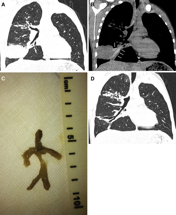
We report the case of a 17-year-old boy with cystic fibrosis (CF) who presented with persistent cough; after starting intravenous antibiotics for he underwent a computed tomography (CT) scan of the chest. CT revealed extensive consolidation in the right lower lobe with relative bronchus obstruction; the cause of bronchial obstruction was detected in the mediastinal window, corresponding to a bronchial tree-shaped, thick, tenacious mucous plug. This was extracted 48 h after unresponsive bronchial washing and endobronchial instillation of rhDNAse, using foreign-body forceps, with subsequent resolution of cough. This case, which is the second report of plastic bronchitis in CF, was resolved by mechanical removal of the mucous plug, suggesting that a careful observation of CT imaging may guide intervention aimed at resolution of atelectasis.
我们报告了一例17岁的囊性纤维化(CF)男孩病例,该男孩持续咳嗽;在开始静脉注射抗生素治疗后,他接受了胸部计算机断层扫描(CT)。CT显示右下叶广泛实变并伴有相对支气管阻塞;在纵隔窗中检测到支气管阻塞的原因,对应于支气管树状、浓稠、坚韧的黏液栓。在支气管冲洗和支气管内滴注重组人脱氧核糖核酸酶(rhDNAse)无效48小时后,使用异物钳取出了该黏液栓,随后咳嗽症状缓解。该病例是CF中塑料支气管炎的第二例报告,通过机械清除黏液栓得以解决,这表明仔细观察CT成像可能会指导旨在解决肺不张的干预措施。