• 文献检索
  • 文档翻译
  • 深度研究
  • 学术资讯
  • Suppr Zotero 插件Zotero 插件
  • 邀请有礼
  • 套餐&价格
  • 历史记录
应用&插件
Suppr Zotero 插件Zotero 插件浏览器插件Mac 客户端Windows 客户端微信小程序
定价
高级版会员购买积分包购买API积分包
服务
文献检索文档翻译深度研究API 文档MCP 服务
关于我们
关于 Suppr公司介绍联系我们用户协议隐私条款
关注我们

Suppr 超能文献

核心技术专利:CN118964589B侵权必究
粤ICP备2023148730 号-1Suppr @ 2026

文献检索

告别复杂PubMed语法,用中文像聊天一样搜索,搜遍4000万医学文献。AI智能推荐,让科研检索更轻松。

立即免费搜索

文件翻译

保留排版,准确专业,支持PDF/Word/PPT等文件格式,支持 12+语言互译。

免费翻译文档

深度研究

AI帮你快速写综述,25分钟生成高质量综述,智能提取关键信息,辅助科研写作。

立即免费体验

基于肝包膜引导超声图像分类的肝硬化诊断方法研究

Learning to Diagnose Cirrhosis with Liver Capsule Guided Ultrasound Image Classification.

机构信息

School of Computer Science, Shanghai Key Laboratory of Intelligent Information Processing, Fudan University, Shanghai 201203, China.

School of Electronic and Electrical Engineering, Shanghai University of Engineering Science, Shanghai 201620, China.

出版信息

Sensors (Basel). 2017 Jan 13;17(1):149. doi: 10.3390/s17010149.

DOI:10.3390/s17010149
PMID:28098774
原文链接:https://pmc.ncbi.nlm.nih.gov/articles/PMC5298722/
Abstract

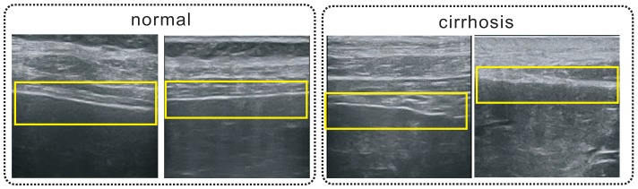
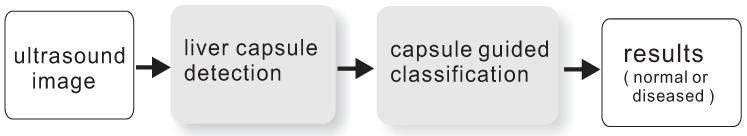
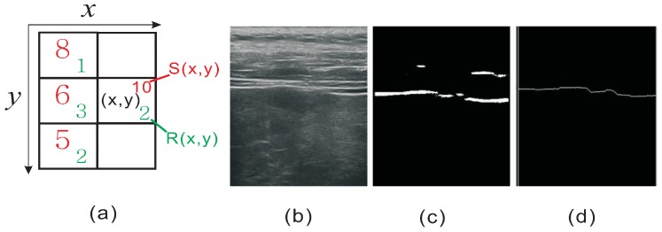
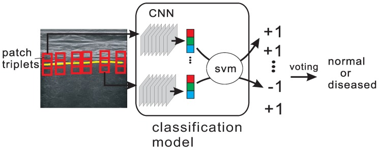
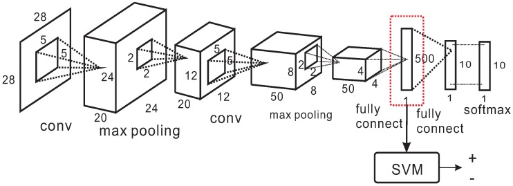
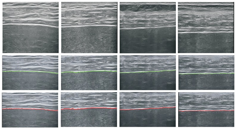
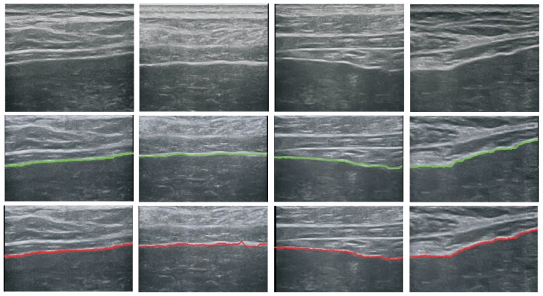
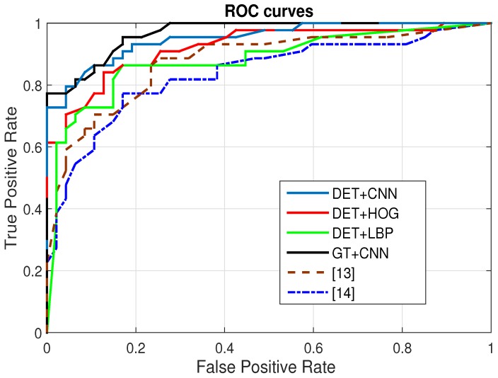
This paper proposes a computer-aided cirrhosis diagnosis system to diagnose cirrhosis based on ultrasound images. We first propose a method to extract a liver capsule on an ultrasound image, then, based on the extracted liver capsule, we fine-tune a deep convolutional neural network (CNN) model to extract features from the image patches cropped around the liver capsules. Finally, a trained support vector machine (SVM) classifier is applied to classify the sample into normal or abnormal cases. Experimental results show that the proposed method can effectively extract the liver capsules and accurately classify the ultrasound images.

摘要

本文提出了一种基于超声图像的计算机辅助肝硬化诊断系统。我们首先提出了一种从超声图像中提取肝包膜的方法,然后,基于提取的肝包膜,我们微调了一个深度卷积神经网络(CNN)模型,从围绕肝包膜裁剪的图像块中提取特征。最后,应用训练好的支持向量机(SVM)分类器对样本进行正常或异常分类。实验结果表明,所提出的方法可以有效地提取肝包膜,并准确地对超声图像进行分类。

https://cdn.ncbi.nlm.nih.gov/pmc/blobs/2fa8/5298722/e0ae7d621ce8/sensors-17-00149-g010.jpg
https://cdn.ncbi.nlm.nih.gov/pmc/blobs/2fa8/5298722/8af3a1c2b796/sensors-17-00149-g001.jpg
https://cdn.ncbi.nlm.nih.gov/pmc/blobs/2fa8/5298722/9881e5f72132/sensors-17-00149-g002.jpg
https://cdn.ncbi.nlm.nih.gov/pmc/blobs/2fa8/5298722/96267895a187/sensors-17-00149-g003.jpg
https://cdn.ncbi.nlm.nih.gov/pmc/blobs/2fa8/5298722/da1b2b70a64d/sensors-17-00149-g004.jpg
https://cdn.ncbi.nlm.nih.gov/pmc/blobs/2fa8/5298722/65e21d02a880/sensors-17-00149-g005.jpg
https://cdn.ncbi.nlm.nih.gov/pmc/blobs/2fa8/5298722/bf9326863da8/sensors-17-00149-g006.jpg
https://cdn.ncbi.nlm.nih.gov/pmc/blobs/2fa8/5298722/c34ff432def7/sensors-17-00149-g007.jpg
https://cdn.ncbi.nlm.nih.gov/pmc/blobs/2fa8/5298722/9218f7b16e5f/sensors-17-00149-g008.jpg
https://cdn.ncbi.nlm.nih.gov/pmc/blobs/2fa8/5298722/1958ce50beff/sensors-17-00149-g009.jpg
https://cdn.ncbi.nlm.nih.gov/pmc/blobs/2fa8/5298722/e0ae7d621ce8/sensors-17-00149-g010.jpg

相似文献

1
Learning to Diagnose Cirrhosis with Liver Capsule Guided Ultrasound Image Classification.基于肝包膜引导超声图像分类的肝硬化诊断方法研究
Sensors (Basel). 2017 Jan 13;17(1):149. doi: 10.3390/s17010149.
2
A novel method for accurate extraction of liver capsule and auxiliary diagnosis of liver cirrhosis based on high-frequency ultrasound images.一种基于高频超声图像准确提取肝包膜及辅助诊断肝硬化的新方法。
Comput Biol Med. 2020 Oct;125:104002. doi: 10.1016/j.compbiomed.2020.104002. Epub 2020 Sep 18.
3
Automated Identification of Hookahs (Waterpipes) on Instagram: An Application in Feature Extraction Using Convolutional Neural Network and Support Vector Machine Classification.Instagram上水烟袋(水烟筒)的自动识别:一种使用卷积神经网络和支持向量机分类进行特征提取的应用。
J Med Internet Res. 2018 Nov 21;20(11):e10513. doi: 10.2196/10513.
4
Preliminary Study of Chronic Liver Classification on Ultrasound Images Using an Ensemble Model.基于集成模型的超声图像慢性肝脏分类初步研究
Ultrason Imaging. 2018 Nov;40(6):357-379. doi: 10.1177/0161734618787447. Epub 2018 Jul 17.
5
Computer assisted recognition of breast cancer in biopsy images via fusion of nucleus-guided deep convolutional features.通过细胞核引导的深度卷积特征融合实现活检图像中乳腺癌的计算机辅助识别。
Comput Methods Programs Biomed. 2020 Oct;194:105531. doi: 10.1016/j.cmpb.2020.105531. Epub 2020 May 11.
6
Decision Fusion-Based Fetal Ultrasound Image Plane Classification Using Convolutional Neural Networks.基于决策融合的卷积神经网络胎儿超声图像平面分类
Ultrasound Med Biol. 2019 May;45(5):1259-1273. doi: 10.1016/j.ultrasmedbio.2018.11.016. Epub 2019 Feb 27.
7
Aggregated micropatch-based deep learning neural network for ultrasonic diagnosis of cirrhosis.基于聚合微补丁的深度学习神经网络用于肝硬化的超声诊断
Artif Intell Med. 2023 May;139:102541. doi: 10.1016/j.artmed.2023.102541. Epub 2023 Mar 31.
8
Computer-aided diagnosis of endobronchial ultrasound images using convolutional neural network.使用卷积神经网络进行支气管内超声图像的计算机辅助诊断。
Comput Methods Programs Biomed. 2019 Aug;177:175-182. doi: 10.1016/j.cmpb.2019.05.020. Epub 2019 May 22.
9
Machine Learning-Aided Chronic Kidney Disease Diagnosis Based on Ultrasound Imaging Integrated with Computer-Extracted Measurable Features.基于超声成像与计算机提取可测量特征融合的机器学习辅助慢性肾脏病诊断。
J Digit Imaging. 2022 Oct;35(5):1091-1100. doi: 10.1007/s10278-022-00625-8. Epub 2022 Apr 11.
10
An Intelligent Diagnosis Method of Brain MRI Tumor Segmentation Using Deep Convolutional Neural Network and SVM Algorithm.基于深度卷积神经网络和支持向量机算法的脑 MRI 肿瘤分割智能诊断方法。
Comput Math Methods Med. 2020 Jul 14;2020:6789306. doi: 10.1155/2020/6789306. eCollection 2020.

引用本文的文献

1
Artificial intelligence-based ultrasound imaging technologies for hepatic diseases.用于肝脏疾病的基于人工智能的超声成像技术。
ILIVER. 2022 Nov 16;1(4):252-264. doi: 10.1016/j.iliver.2022.11.001. eCollection 2022 Dec.
2
Bibliometrics of artificial intelligence applications in hepatobiliary surgery from 2014 to 2024.2014年至2024年人工智能在肝胆外科应用的文献计量学
World J Gastrointest Surg. 2025 May 27;17(5):104728. doi: 10.4240/wjgs.v17.i5.104728.
3
Explainable Artificial Intelligence for Diagnosis and Staging of Liver Cirrhosis Using Stacked Ensemble and Multi-Task Learning.

本文引用的文献

1
Artificial neural network application in the diagnosis of disease conditions with liver ultrasound images.人工神经网络在利用肝脏超声图像诊断疾病状况中的应用。
Adv Bioinformatics. 2014;2014:708279. doi: 10.1155/2014/708279. Epub 2014 Sep 16.
2
Evolution-based hierarchical feature fusion for ultrasonic liver tissue characterization.基于进化的层次特征融合用于超声肝脏组织表征
IEEE J Biomed Health Inform. 2013 Sep;17(5):967-76. doi: 10.1109/JBHI.2013.2261819.
3
SVM-based characterization of liver ultrasound images using wavelet packet texture descriptors.
使用堆叠集成和多任务学习的可解释人工智能用于肝硬化的诊断和分期
Diagnostics (Basel). 2025 May 6;15(9):1177. doi: 10.3390/diagnostics15091177.
4
Advances in Deep Learning-Based Medical Image Analysis.基于深度学习的医学图像分析进展
Health Data Sci. 2021 May 19;2021:8786793. doi: 10.34133/2021/8786793. eCollection 2021.
5
Quantitative methods for optimizing patient outcomes in liver transplantation.肝移植中优化患者预后的定量方法。
Liver Transpl. 2024 Mar 1;30(3):311-320. doi: 10.1097/LVT.0000000000000325. Epub 2023 Dec 25.
6
Improving nonalcoholic fatty liver disease classification performance with latent diffusion models.利用潜在扩散模型提高非酒精性脂肪性肝病分类性能。
Sci Rep. 2023 Dec 7;13(1):21619. doi: 10.1038/s41598-023-48062-z.
7
Radiological Diagnosis of Chronic Liver Disease and Hepatocellular Carcinoma: A Review.慢性肝病和肝细胞癌的放射学诊断:综述。
J Med Syst. 2023 Jul 11;47(1):73. doi: 10.1007/s10916-023-01968-7.
8
Hepatocellular Carcinoma Recognition from Ultrasound Images Using Combinations of Conventional and Deep Learning Techniques.基于常规与深度学习技术联合的超声图像肝细胞癌识别。
Sensors (Basel). 2023 Feb 24;23(5):2520. doi: 10.3390/s23052520.
9
Automatic Detection and Measurement of Renal Cysts in Ultrasound Images: A Deep Learning Approach.超声图像中肾囊肿的自动检测与测量:一种深度学习方法。
Healthcare (Basel). 2023 Feb 7;11(4):484. doi: 10.3390/healthcare11040484.
10
Noninvasive Assessment of Liver Fibrosis and Inflammation in Chronic Hepatitis B: A Dual-task Convolutional Neural Network (DtCNN) Model Based on Ultrasound Shear Wave Elastography.慢性乙型肝炎中肝纤维化和炎症的无创评估:基于超声剪切波弹性成像的双任务卷积神经网络(DtCNN)模型
J Clin Transl Hepatol. 2022 Dec 28;10(6):1077-1085. doi: 10.14218/JCTH.2021.00447. Epub 2022 Mar 29.
基于支持向量机的肝超声图像小波包纹理特征描述
J Digit Imaging. 2013 Jun;26(3):530-43. doi: 10.1007/s10278-012-9537-8.
4
Non-invasive evaluation of liver cirrhosis using ultrasound.使用超声对肝硬化进行无创评估。
Clin Radiol. 2009 Nov;64(11):1056-66. doi: 10.1016/j.crad.2009.05.010. Epub 2009 Aug 14.
5
Texture features for classification of ultrasonic liver images.超声肝脏图像分类的纹理特征。
IEEE Trans Med Imaging. 1992;11(2):141-52. doi: 10.1109/42.141636.
6
A texture classification method for diffused liver diseases using Gabor wavelets.一种使用伽柏小波的弥漫性肝病纹理分类方法。
Conf Proc IEEE Eng Med Biol Soc. 2005;2005:1567-70. doi: 10.1109/IEMBS.2005.1616734.
7
Ultrasonic liver tissues classification by fractal feature vector based on M-band wavelet transform.基于M带小波变换的分形特征向量对肝脏组织进行超声分类
IEEE Trans Med Imaging. 2003 Mar;22(3):382-92. doi: 10.1109/TMI.2003.809593.
8
Characterization of visually similar diffuse diseases from B-scan liver images using nonseparable wavelet transform.使用不可分离小波变换从肝脏B超图像中鉴别视觉上相似的弥漫性疾病
IEEE Trans Med Imaging. 1998 Aug;17(4):541-9. doi: 10.1109/42.730399.
9
Quantitative ultrasonic detection and classification of diffuse liver disease. Comparison with human observer performance.弥漫性肝病的定量超声检测与分类。与人类观察者表现的比较。
Invest Radiol. 1989 Mar;24(3):196-203. doi: 10.1097/00004424-198903000-00004.
10
Cirrhosis: diagnosis with sonographic study of the liver surface.肝硬化:通过肝脏表面超声检查进行诊断。
Radiology. 1989 Aug;172(2):389-92. doi: 10.1148/radiology.172.2.2526349.