Barnes Laurent, Saurat Jean-Hilaire, Kaya Gürkan
Department of Dermatology, University Hospital of Geneva, University of Geneva, Geneva, Switzerland.
Swiss Center for Applied Human Toxicology, University of Geneva, Geneva, Switzerland.
PLoS One. 2017 Jan 18;12(1):e0169452. doi: 10.1371/journal.pone.0169452. eCollection 2017.
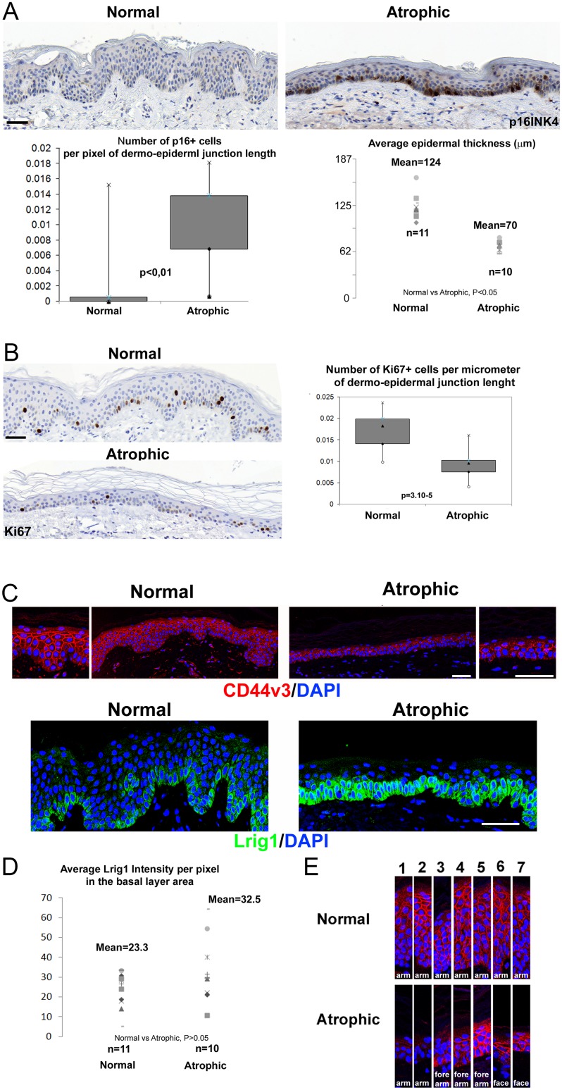
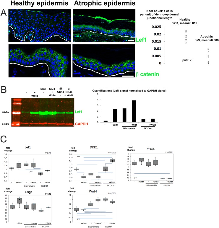
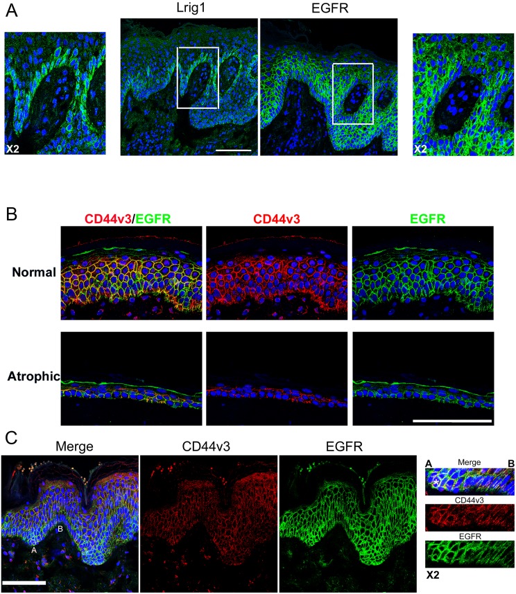
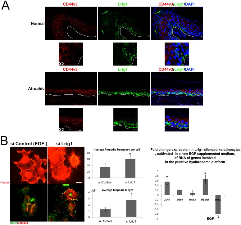
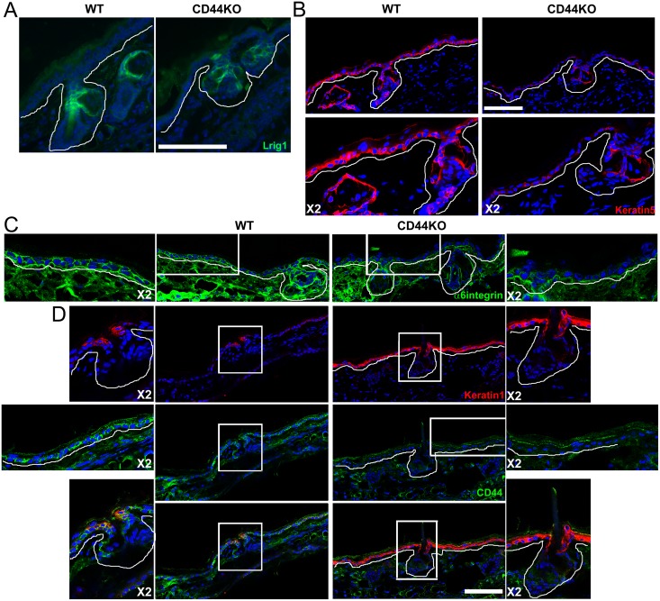
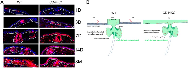
Lrig1 is known to repress the epidermal growth through its inhibitory activity on EGFR, while CD44 promotes it. We analyzed the expression of these molecules in senescent atrophic human epidermis and in the epidermis of CD44KO mice. In normal human epidermis, Lrig1+ cells form clusters located in the basal layer in which CD44 expression is downregulated and Lef1 expression reflects an active Wnt signaling. In senescent atrophic human epidermis, we found retention of Lrig1high+ cells all along the basal layer, forming no clusters, with decrease of CD44 and lef1 expression. In vitro silencing of CD44 indicated that CD44 may be required for Wnt signaling. However, if looking at the ear epidermis of CD44KO mice, we only found a limited interfollicular epidermal atrophy and unchanged Lrig1high+ cells in the hair follicle. Cell lineage tracing further revealed that interfollicular epidermis did lost its self-renewing capacity but that its homeostasis relied on Lrig1-derived keratinocytes migrating from the hair follicle. Therefore, we conclude that CD44 downregulation is part of the phenotype of senescent atrophic human epidermis, and contributes to reduce Wnt signaling and to alter Lrig1high+ stem cell distribution.
已知Lrig1通过其对表皮生长因子受体(EGFR)的抑制活性来抑制表皮生长,而CD44则促进表皮生长。我们分析了这些分子在衰老萎缩的人类表皮和CD44基因敲除(CD44KO)小鼠的表皮中的表达情况。在正常人类表皮中,Lrig1阳性细胞形成位于基底层的簇,其中CD44表达下调,而淋巴样增强因子1(Lef1)表达反映活跃的Wnt信号传导。在衰老萎缩的人类表皮中,我们发现Lrig1高表达阳性细胞沿基底层全程保留,不形成簇,CD44和Lef1表达降低。体外沉默CD44表明Wnt信号传导可能需要CD44。然而,观察CD44KO小鼠的耳部表皮,我们仅发现有限的毛囊间表皮萎缩以及毛囊中Lrig1高表达阳性细胞未改变。细胞谱系追踪进一步显示,毛囊间表皮确实丧失了自我更新能力,但其稳态依赖于从毛囊迁移而来的Lrig1来源的角质形成细胞。因此,我们得出结论,CD44下调是衰老萎缩人类表皮表型的一部分,并有助于减少Wnt信号传导和改变Lrig1高表达阳性干细胞分布。