Speisky Daniela, de Davila María Teresa García, Vigovich Felix, Mendez Julian, Maurette Rafael, Ejarque Marcos García, Spina Juan Carlos, Iotti Alejandro, Dezanzo Pablo
Department of Histopathology, Hospital Británico, Buenos Aires C1280AEB, Argentina.
Department of Hepatobiliary Surgery, Hospital Británico, Buenos Aires C1280AEB, Argentina.
Ecancermedicalscience. 2016 Nov 22;10:693. doi: 10.3332/ecancer.2016.693. eCollection 2016.
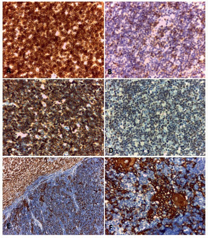
Thymomas are rare tumours characterised by their slow growth and capacity to invade directly by contiguity. While distant dissemination is infrequent, all sub-types of thymoma have the capacity to metastasise to extrathoracic organs. We present here the case of a female patient with a liver mass discovered 13 years after the removal of a mediastinal thymoma and after ten years from thyroidectomy for papillary carcinoma. The histopathological study showed that the lesion contained an epithelial component, which was immunohistochemically positive for pankeratin. It was accompanied by numerous small lymphocytes testing positive for TdT, CD3, CD4, CD5, CD8, CD99, and CD43. The result was consistent with hepatic metastatic thymoma sub-type B1, according to the World Health Organisation classification (WHO). Our case highlights the importance of morphological and immunohistological examinations in the differential diagnosis of visceral masses in patients with a history of thymoma. Given the infrequency of its metastasis and the increased risk of developing other primary tumours that these patients have, these studies play a significant role.
胸腺瘤是一种罕见的肿瘤,其特点是生长缓慢且有直接连续性侵袭的能力。虽然远处播散不常见,但所有胸腺瘤亚型都有转移至胸外器官的能力。我们在此介绍一例女性患者,该患者在纵隔胸腺瘤切除13年后以及甲状腺乳头状癌甲状腺切除术后10年发现肝脏肿物。组织病理学研究显示,该病变包含上皮成分,免疫组化检测显示其细胞角蛋白呈阳性。同时伴有大量小淋巴细胞,末端脱氧核苷酸转移酶(TdT)、CD3、CD4、CD5、CD8、CD99和CD43检测呈阳性。根据世界卫生组织(WHO)分类,结果符合B1型肝转移性胸腺瘤。我们的病例突出了形态学和免疫组织学检查在有胸腺瘤病史患者内脏肿物鉴别诊断中的重要性。鉴于其转移罕见,且这些患者发生其他原发性肿瘤的风险增加,这些研究具有重要意义。