Maanja Maren, Wieslander Björn, Schlegel Todd T, Bacharova Ljuba, Abu Daya Hussein, Fridman Yaron, Wong Timothy C, Schelbert Erik B, Ugander Martin
Department of Clinical Physiology, Karolinska Institutet, and Karolinska University Hospital, Stockholm, Sweden.
Department of Medicine, University of Pittsburgh Medical Center, Pittsburgh, PA.
J Am Heart Assoc. 2017 Jan 22;6(1):e003795. doi: 10.1161/JAHA.116.003795.
Myocardial fibrosis quantified by myocardial extracellular volume fraction (ECV) and left ventricular mass (LVM) index (LVMI) measured by cardiovascular magnetic resonance might represent independent and opposing contributors to ECG voltage measures of left ventricular hypertrophy (LVH). Diffuse myocardial fibrosis can occur in LVH and interfere with ECG voltage measures. This phenomenon could explain the decreased sensitivity of LVH detectable by ECG, a fundamental diagnostic tool in cardiology.
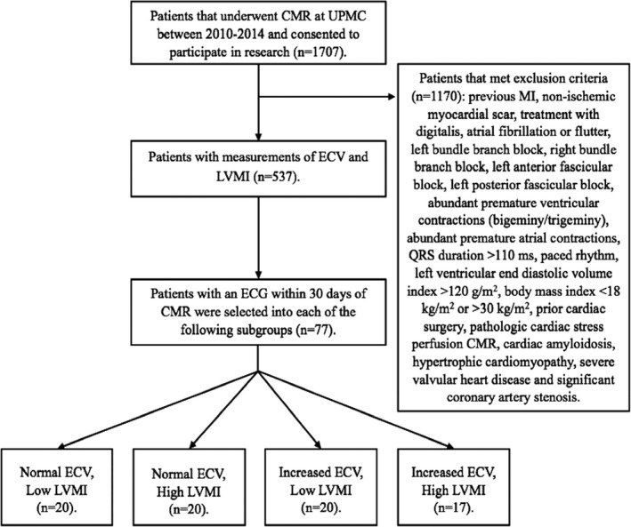
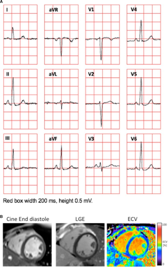
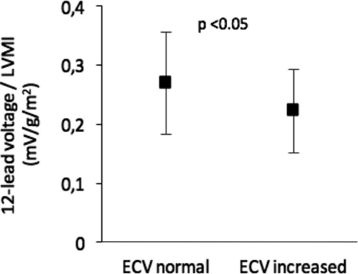
We identified 77 patients (median age, 53 [interquartile range, 26-60] years; 49% female) referred for contrast-enhanced cardiovascular magnetic resonance with ECV measures and 12-lead ECG. Exclusion criteria included clinical confounders that might influence ECG measures of LVH. We evaluated ECG voltage-based LVH measures, including Sokolow-Lyon index, Cornell voltage, 12-lead voltage, and the vectorcardiogram spatial QRS voltage, with respect to LVMI and ECV. ECV and LVMI were not correlated (R=0.02; P=0.25). For all voltage-related parameters, higher LVMI resulted in greater voltage (r=0.33-0.49; P<0.05 for all), whereas increased ECV resulted in lower voltage (r=-0.32 to -0.57; P<0.05 for all). When accounting for body fat, LV end-diastolic volume, and mass-to-volume ratio, both LVMI (β=0.58, P=0.03) and ECV (β=-0.46, P<0.001) were independent predictors of QRS voltage (multivariate adjusted R=0.39; P<0.001).
Myocardial mass and diffuse myocardial fibrosis have independent and opposing effects upon ECG voltage measures of LVH. Diffuse myocardial fibrosis quantified by ECV can obscure the ECG manifestations of increased LVM. This provides mechanistic insight, which can explain the limited sensitivity of the ECG for detecting increased LVM.
通过心血管磁共振测量的心肌细胞外容积分数(ECV)和左心室质量(LVM)指数(LVMI)量化的心肌纤维化,可能分别独立且相反地影响左心室肥厚(LVH)心电图电压测量结果。弥漫性心肌纤维化可发生于LVH,并干扰心电图电压测量。这种现象可以解释心电图作为心脏病学基本诊断工具检测LVH敏感度下降的原因。
我们纳入了77例接受对比增强心血管磁共振ECV测量及12导联心电图检查的患者(年龄中位数53岁[四分位间距26 - 60岁];49%为女性)。排除标准包括可能影响LVH心电图测量结果的临床混杂因素。我们评估了基于心电图电压的LVH测量指标,包括索科洛夫 - 里昂指数、康奈尔电压、12导联电压和向量心电图空间QRS电压,与LVMI和ECV的关系。ECV与LVMI不相关(R = 0.02;P = 0.25)。对于所有与电压相关的参数,LVMI越高,电压越高(r = 0.33 - 0.49;所有P < 0.05),而ECV升高则导致电压降低(r = -0.32至 -0.57;所有P < 0.05)。在考虑体脂、左心室舒张末期容积和质量体积比时,LVMI(β = 0.58,P = 0.03)和ECV(β = -0.46,P < 0.001)均为QRS电压的独立预测因素(多变量调整R = 0.39;P < 0.001)。
心肌质量和弥漫性心肌纤维化对LVH心电图电压测量具有独立且相反的影响。通过ECV量化的弥漫性心肌纤维化可掩盖LVM增加的心电图表现。这提供了机制性见解,可解释心电图检测LVM增加的敏感性有限的原因。