Ehrenberg A J, Nguy A K, Theofilas P, Dunlop S, Suemoto C K, Di Lorenzo Alho A T, Leite R P, Diehl Rodriguez R, Mejia M B, Rüb U, Farfel J M, de Lucena Ferretti-Rebustini R E, Nascimento C F, Nitrini R, Pasquallucci C A, Jacob-Filho W, Miller B, Seeley W W, Heinsen H, Grinberg L T
University of California, San Francisco, California, USA.
University of California, Berkeley, California, USA.
Neuropathol Appl Neurobiol. 2017 Aug;43(5):393-408. doi: 10.1111/nan.12387. Epub 2017 Mar 31.
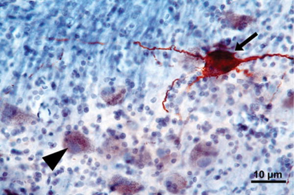
Hyperphosphorylated tau neuronal cytoplasmic inclusions (ht-NCI) are the best protein correlate of clinical decline in Alzheimer's disease (AD). Qualitative evidence identifies ht-NCI accumulating in the isodendritic core before the entorhinal cortex. Here, we used unbiased stereology to quantify ht-NCI burden in the locus coeruleus (LC) and dorsal raphe nucleus (DRN), aiming to characterize the impact of AD pathology in these nuclei with a focus on early stages.
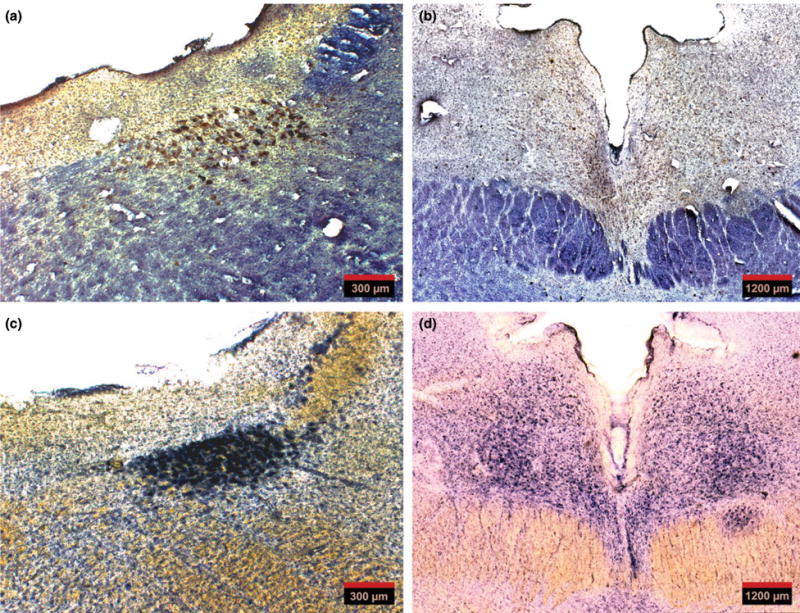
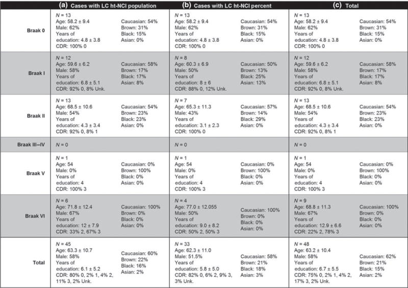
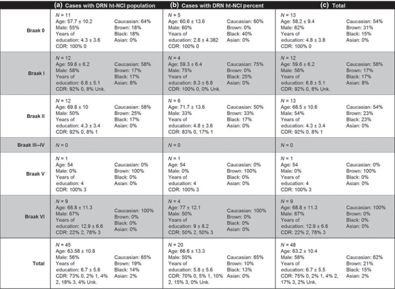
We utilized unbiased stereology in a sample of 48 well-characterized subjects enriched for controls and early AD stages. ht-NCI counts were estimated in 60-μm-thick sections immunostained for p-tau throughout LC and DRN. Data were integrated with unbiased estimates of LC and DRN neuronal population for a subset of cases.
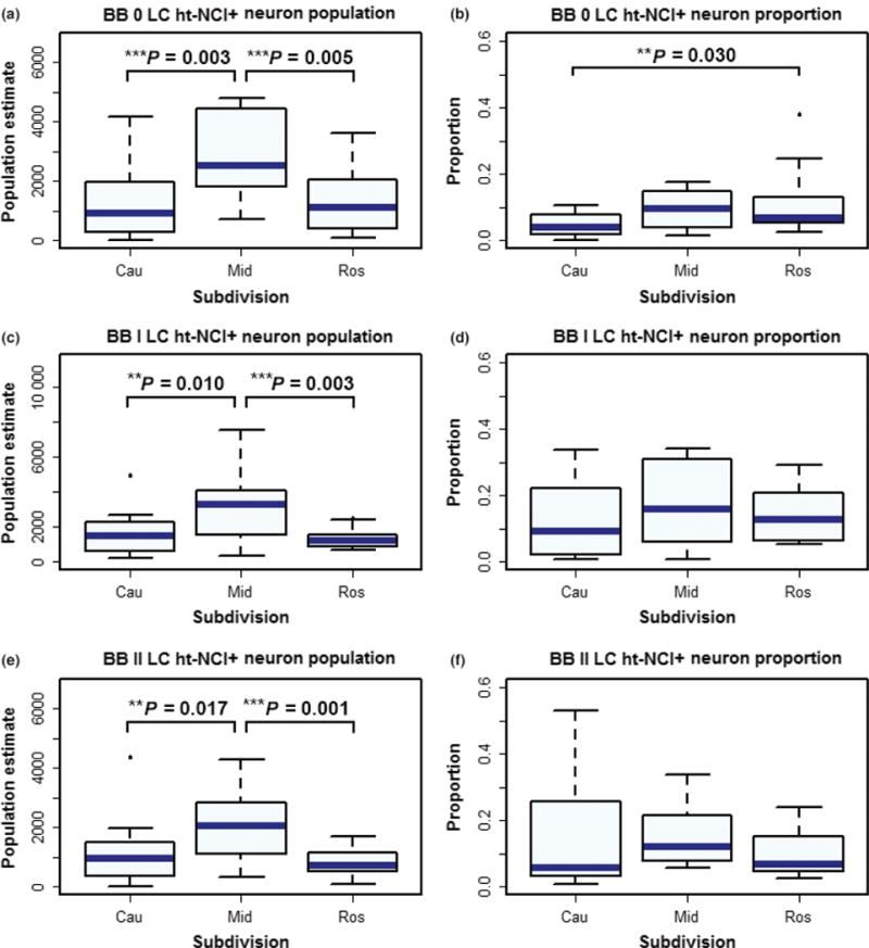
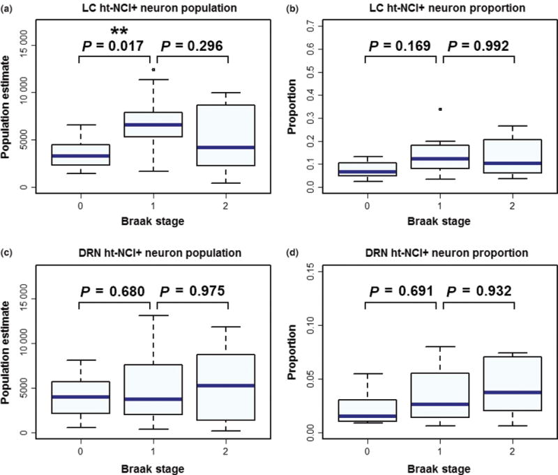
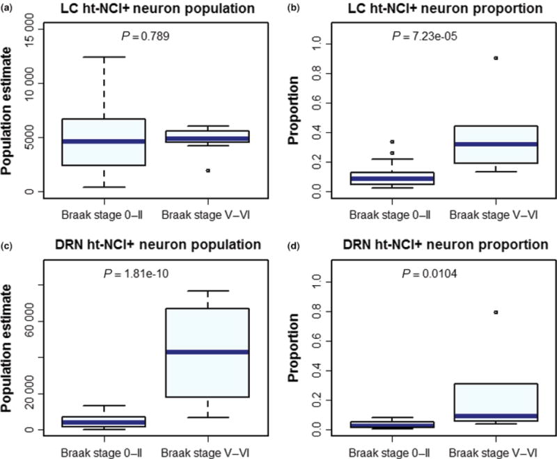
In Braak stage 0, 7.9% and 2.6% of neurons in LC and DRN, respectively, harbour ht-NCIs. Although the number of ht-NCI+ neurons significantly increased by about 1.9× between Braak stages 0 to I in LC (P = 0.02), we failed to detect any significant difference between Braak stage I and II. Also, the number of ht-NCI+ neurons remained stable in DRN between all stages 0 and II. Finally, the differential susceptibility to tau inclusions among nuclear subdivisions was more notable in LC than in DRN.
LC and DRN neurons exhibited ht-NCI during AD precortical stages. The ht-NCI increases along AD progression on both nuclei, but quantitative changes in LC precede DRN changes.
高磷酸化tau蛋白神经元胞质包涵体(ht-NCI)是阿尔茨海默病(AD)临床衰退的最佳蛋白质相关指标。定性证据表明ht-NCI在内嗅皮质之前在等树突核心中积累。在此,我们使用无偏倚立体学方法来量化蓝斑(LC)和中缝背核(DRN)中ht-NCI的负担,旨在表征AD病理在这些核中的影响,重点关注早期阶段。
我们在48名特征明确的受试者样本中运用无偏倚立体学方法,这些受试者以对照和AD早期阶段为主。在整个LC和DRN中对p-tau进行免疫染色的60μm厚切片中估计ht-NCI计数。对于一部分病例,数据与LC和DRN神经元群体的无偏估计值相结合。
在Braak 0期,LC和DRN中分别有7.9%和2.6%的神经元含有ht-NCI。尽管在LC中,从Braak 0期到I期,ht-NCI+神经元的数量显著增加了约1.9倍(P = 0.02),但我们未能检测到Braak I期和II期之间有任何显著差异。此外,在DRN中,所有0期到II期之间ht-NCI+神经元的数量保持稳定。最后,核亚区对tau包涵体的易感性差异在LC中比在DRN中更明显。
在AD皮质前阶段,LC和DRN神经元表现出ht-NCI。随着AD进展,两个核中的ht-NCI都增加,但LC中的定量变化先于DRN的变化。