Dichtel Laura E, Corey Kathleen E, Misdraji Joseph, Bredella Miriam A, Schorr Melanie, Osganian Stephanie A, Young Brian J, Sung Joshua C, Miller Karen K
Neuroendocrine Unit, Massachusetts General Hospital/Harvard Medical School, Boston, Massachusetts, USA.
Division of Gastroenterology, Massachusetts General Hospital/Harvard Medical School, Boston, Massachusetts, USA.
Clin Transl Gastroenterol. 2017 Jan 26;8(1):e217. doi: 10.1038/ctg.2016.72.
The mechanisms responsible for the development of nonalcoholic fatty liver disease (NAFLD) and progression to nonalcoholic steatohepatitis (NASH) are incompletely understood. Growing evidence suggests that growth hormone (GH) and insulin-like growth factor-1 (IGF-1) may have roles in the development and progression of NAFLD. We hypothesized that lower serum IGF-1 levels would be associated with increased liver fat accumulation, inflammation, and fibrosis in a group of meticulously phenotyped obese subjects with liver biopsies.
A retrospective, cross-sectional study was performed at Massachusetts General Hospital, Boston, MA, USA and St. Mary's Hospital, Richmond, VA, USA. Liver biopsies were performed in 142 subjects during NAFLD work-up or bariatric surgery and were graded by a single, blinded pathologist. Main outcome measures included liver histology and serum IGF-1.
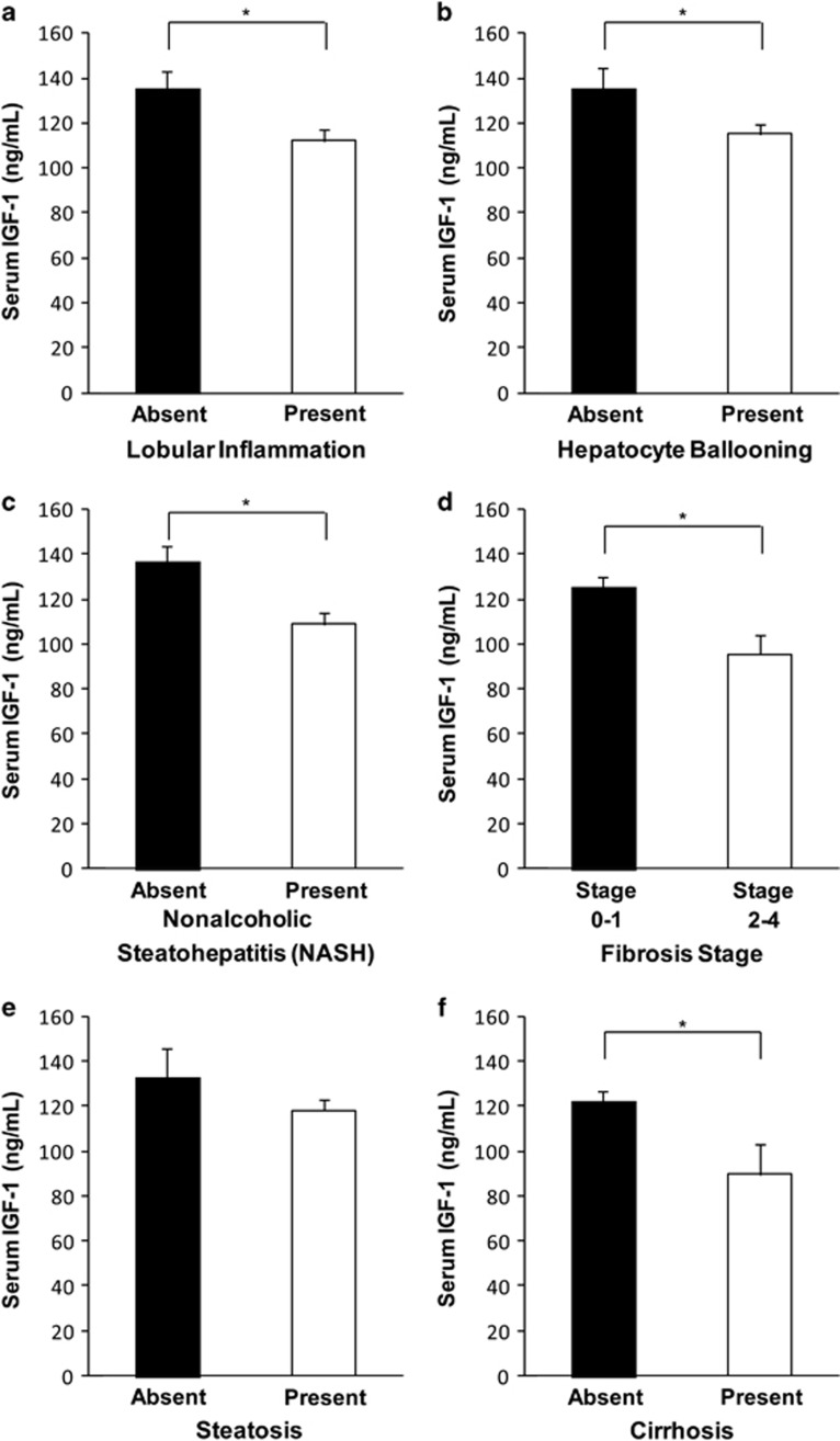
Mean age was 52±10 years and body mass index (BMI) was 43±9 kg/m. Mean serum IGF-1 was lower in subjects with lobular inflammation (112±47 vs. 136±57 ng/ml, P=0.01), hepatocyte ballooning (115±48 vs. 135±57 ng/ml, P=0.05), higher fibrosis stage (stage 2-4 vs. 0-1; 96±40 vs. 125±51 ng/ml, P=0.005), and NASH (109±45 vs. 136±57 ng/ml, P=0.002). All results remained significant after controlling for age, BMI, and a diagnosis of diabetes, and all but hepatocyte ballooning (trend, P=0.06) remained significant after excluding individuals with cirrhosis. Steatosis was not significantly associated with mean serum IGF-1 levels.
Low serum IGF-1 levels are associated with increased histologic severity of NAFLD when rigorously controlled for age, BMI, the presence of diabetes, and after the exclusion of subjects with cirrhosis. Further investigation is warranted to determine the differential effects of GH and IGF-1 on the development and progression of NAFLD, which could further elucidate pathophysiology and identify therapeutic targets.
非酒精性脂肪性肝病(NAFLD)的发病机制以及其向非酒精性脂肪性肝炎(NASH)进展的机制尚未完全明确。越来越多的证据表明,生长激素(GH)和胰岛素样生长因子-1(IGF-1)可能在NAFLD的发生和发展中起作用。我们推测,在一组经过细致表型分析且接受了肝活检的肥胖受试者中,较低的血清IGF-1水平将与肝脏脂肪堆积增加、炎症和纤维化相关。
在美国马萨诸塞州波士顿的麻省总医院和美国弗吉尼亚州里士满的圣玛丽医院进行了一项回顾性横断面研究。在142名受试者进行NAFLD检查或减肥手术期间进行了肝活检,并由一名单盲病理学家进行分级。主要观察指标包括肝脏组织学和血清IGF-1。
平均年龄为52±10岁,体重指数(BMI)为43±9kg/m²。小叶炎症患者(112±47对136±57ng/ml,P=0.01)、肝细胞气球样变患者(115±48对135±57ng/ml,P=0.05)、纤维化程度较高阶段(2-4期对0-1期;96±40对125±51ng/ml,P=0.005)以及NASH患者(109±45对136±57ng/ml,P=0.002)的平均血清IGF-1水平较低。在控制年龄、BMI和糖尿病诊断后,所有结果仍然显著,并且在排除肝硬化患者后,除肝细胞气球样变外(趋势,P=0.06)所有结果仍然显著。脂肪变性与平均血清IGF-1水平无显著相关性。
在严格控制年龄、BMI、糖尿病存在情况以及排除肝硬化患者后,低血清IGF-1水平与NAFLD组织学严重程度增加相关。有必要进一步研究以确定GH和IGF-1对NAFLD发生和发展的不同影响,这可能进一步阐明病理生理学并确定治疗靶点。