• 文献检索
  • 文档翻译
  • 深度研究
  • 学术资讯
  • Suppr Zotero 插件Zotero 插件
  • 邀请有礼
  • 套餐&价格
  • 历史记录
应用&插件
Suppr Zotero 插件Zotero 插件浏览器插件Mac 客户端Windows 客户端微信小程序
定价
高级版会员购买积分包购买API积分包
服务
文献检索文档翻译深度研究API 文档MCP 服务
关于我们
关于 Suppr公司介绍联系我们用户协议隐私条款
关注我们

Suppr 超能文献

核心技术专利:CN118964589B侵权必究
粤ICP备2023148730 号-1Suppr @ 2026

文献检索

告别复杂PubMed语法,用中文像聊天一样搜索,搜遍4000万医学文献。AI智能推荐,让科研检索更轻松。

立即免费搜索

文件翻译

保留排版,准确专业,支持PDF/Word/PPT等文件格式,支持 12+语言互译。

免费翻译文档

深度研究

AI帮你快速写综述,25分钟生成高质量综述,智能提取关键信息,辅助科研写作。

立即免费体验

髋臼发育不良相关放射学表现的多平面评估及其在亚洲人群中的患病率调查:一项基于CT的研究

Multiplanar evaluation of radiological findings associated with acetabular dysplasia and investigation of its prevalence in an Asian population: a CT-based study.

作者信息

Mimura Tomohiro, Mori Kanji, Kitagawa Masahiro, Ueki Mariko, Furuya Yuki, Kawasaki Taku, Imai Shinji

机构信息

Department of Orthopedic Surgery, Shiga University of Medical Science, Tsukinowa-cho, Seta, Otsu, Shiga, 520-2192, Japan.

出版信息

BMC Musculoskelet Disord. 2017 Jan 31;18(1):50. doi: 10.1186/s12891-017-1426-3.

DOI:10.1186/s12891-017-1426-3
PMID:28137249
原文链接:https://pmc.ncbi.nlm.nih.gov/articles/PMC5282645/
Abstract

BACKGROUND

Acetabular dysplasia (AD) is a well-known cause of osteoarthritis (OA) of the hip, with its prevalence previously determined on plain radiography. The prevalence of preexisting AD was reported as 7.3% in a patient-based Asian population. Although computed tomography (CT) could evaluate AD in multiple planes, its prevalence using multiplanar CT images has not been reported. We investigated its prevalence with CT on coronal, axial, and sagittal planes and then determined if adding the axial and sagittal planes enhanced the investigation.

METHODS

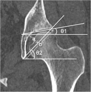
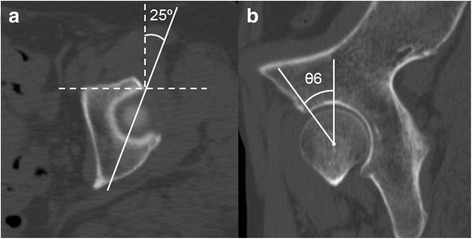
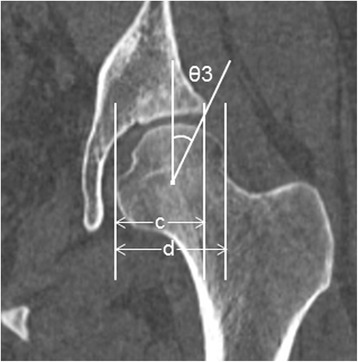
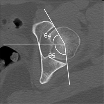
We retrospectively examined 52 consecutive Japanese individuals (mean age 59.4 years) who had undergone CT for conditions unrelated to hip disorders. The inclusion criteria of CT images were (1) reconstructed axial slice thickness of ≤1 mm and (2) normal pelvic rotations and tilt. Exclusion criteria were (1) age <20 years, (2) neither hip center could be clearly detected, (3) evidence of hip OA. The parameters used to define AD on the coronal plane were the center-edge angle, Sharp angle, acetabular index, acetabular depth ratio, and acetabulum head index. The anterior and posterior acetabular sector angles were used as axial parameters and the vertical-center-anterior margin angle as the sagittal parameter. AD prevalence was calculated using multiplanar images and then compared with the previously reported Asian prevalence using 95% confidence intervals (CI). In this study, we defined "prevalence" as the proportion of subjects who had AD in at least one hip.

RESULTS

The mean prevalence of AD on coronal, axial, and sagittal planes was 16.9, 15.4, and 7.7%, respectively. The lowest prevalence found by combining the three planes was 25.0% (95% CI 15.2-38.2%). This prevalence was significantly higher than that in the previously reported Asian population (7.3%).

CONCLUSIONS

At the lowest estimate, the prevalence of AD evaluated in three planes was more than twice as high as the previously reported prevalence in Asians when we investigated its prevalence using multiplanar images. The prevalence of AD in the axial and sagittal planes was not negligible. We therefore suggest that it is important to add axial and sagittal planes' data when investigating the prevalence of AD.

摘要

背景

髋臼发育不良(AD)是髋关节骨关节炎(OA)的一个众所周知的病因,其患病率此前通过X线平片确定。在一个以患者为基础的亚洲人群中,既往AD的患病率报告为7.3%。尽管计算机断层扫描(CT)可以在多个平面评估AD,但其在多平面CT图像上的患病率尚未见报道。我们通过CT在冠状面、轴位和矢状面上研究其患病率,然后确定增加轴位和矢状面是否能加强研究。

方法

我们回顾性检查了52例连续的日本个体(平均年龄59.4岁),他们因与髋关节疾病无关的情况接受了CT检查。CT图像的纳入标准为:(1)重建轴位切片厚度≤1mm;(2)骨盆旋转和倾斜正常。排除标准为:(1)年龄<20岁;(2)双侧髋关节中心均无法清晰检测到;(3)髋关节OA的证据。用于在冠状面定义AD的参数为中心边缘角、夏普角、髋臼指数、髋臼深度比和髋臼头指数。髋臼前后扇形角用作轴位参数,垂直中心前缘角用作矢状位参数。使用多平面图像计算AD患病率,然后使用95%置信区间(CI)与先前报道的亚洲患病率进行比较。在本研究中,我们将“患病率”定义为至少一侧髋关节患有AD的受试者比例。

结果

AD在冠状面、轴位和矢状面的平均患病率分别为16.9%、15.4%和7.7%。三个平面联合发现的最低患病率为25.0%(95%CI 15.2 - 38.2%)。该患病率显著高于先前报道的亚洲人群(7.3%)。

结论

在我们使用多平面图像研究AD患病率时,最低估计值显示,在三个平面评估的AD患病率是先前报道的亚洲人患病率的两倍多。AD在轴位和矢状面的患病率不可忽视。因此,我们建议在调查AD患病率时增加轴位和矢状面的数据很重要。

https://cdn.ncbi.nlm.nih.gov/pmc/blobs/2fcf/5282645/0569f4c25e9f/12891_2017_1426_Fig4_HTML.jpg
https://cdn.ncbi.nlm.nih.gov/pmc/blobs/2fcf/5282645/6a7e59b8ae03/12891_2017_1426_Fig1_HTML.jpg
https://cdn.ncbi.nlm.nih.gov/pmc/blobs/2fcf/5282645/d4396fdde22c/12891_2017_1426_Fig2_HTML.jpg
https://cdn.ncbi.nlm.nih.gov/pmc/blobs/2fcf/5282645/54c05c141499/12891_2017_1426_Fig3_HTML.jpg
https://cdn.ncbi.nlm.nih.gov/pmc/blobs/2fcf/5282645/0569f4c25e9f/12891_2017_1426_Fig4_HTML.jpg

相似文献

1
Multiplanar evaluation of radiological findings associated with acetabular dysplasia and investigation of its prevalence in an Asian population: a CT-based study.髋臼发育不良相关放射学表现的多平面评估及其在亚洲人群中的患病率调查:一项基于CT的研究
BMC Musculoskelet Disord. 2017 Jan 31;18(1):50. doi: 10.1186/s12891-017-1426-3.
2
Tilt-adjusted Cup Anteversion in Patients with Severe Backward Pelvic Tilt is Associated with the Risk of Iliopsoas Impingement: A Three-dimensional Implantation Simulation.倾斜调整杯前倾角在严重后倾骨盆患者中与髂腰肌撞击风险相关:三维植入模拟。
Clin Orthop Relat Res. 2019 Oct;477(10):2243-2254. doi: 10.1097/CORR.0000000000000830.
3
Does Acetabular Coverage Vary Between the Supine and Standing Positions in Patients with Hip Dysplasia?髋臼覆盖在髋关节发育不良患者的仰卧位和站立位之间是否不同?
Clin Orthop Relat Res. 2019 Nov;477(11):2455-2466. doi: 10.1097/CORR.0000000000000898.
4
Hip dysplasia is more severe in Charcot-Marie-Tooth disease than in developmental dysplasia of the hip.髋关节发育不良在夏科-马里-图思病中比在发育性髋关节发育不良中更为严重。
Clin Orthop Relat Res. 2014 Feb;472(2):665-73. doi: 10.1007/s11999-013-3127-z.
5
Effect of coronal plane acetabular correction on joint contact pressure in Periacetabular osteotomy: a finite-element analysis.髋臼冠状面矫正对髋臼周围截骨术关节接触压力的影响:有限元分析。
BMC Musculoskelet Disord. 2022 Jan 14;23(1):48. doi: 10.1186/s12891-022-05005-5.
6
Focal concavity of posterior superior acetabulum and its relation with acetabular dysplasia and retroversion in adults without advanced hip osteoarthritis.成人后上髋臼的局灶性凹陷及其与髋臼发育不良和髋臼后倾的关系,无晚期髋骨关节炎。
BMC Musculoskelet Disord. 2015 Nov 2;16:330. doi: 10.1186/s12891-015-0791-z.
7
Hip morphologic measurements in an Egyptian population.埃及人群的髋部形态学测量
Orthopedics. 2011 Apr 11;34(4). doi: 10.3928/01477447-20110228-10.
8
Prevalence of radiological femoroacetabular impingement in Japanese hip joints: detailed investigation with computed tomography.日本髋关节放射学股骨髋臼撞击症的患病率:计算机断层扫描详细调查
J Orthop Sci. 2015 Jul;20(4):649-56. doi: 10.1007/s00776-015-0733-5. Epub 2015 May 12.
9
Correlation of tilt of the anterior pelvic plane angle with anatomical pelvic tilt and morphological configuration of the acetabulum in patients with developmental dysplasia of the hip: a cross-sectional study.骨盆倾斜角前倾平面与解剖性骨盆倾斜和髋臼形态结构在发育性髋关节发育不良患者中的相关性:一项横断面研究。
J Orthop Surg Res. 2019 Oct 17;14(1):323. doi: 10.1186/s13018-019-1382-8.
10
Anterior centre-edge angle on sagittal CT: a comparison of normal hips to dysplastic hips.矢状面CT上的前中心边缘角:正常髋关节与发育不良髋关节的比较。
Hip Int. 2018 Sep;28(5):535-541. doi: 10.1177/1120700017752569. Epub 2018 May 17.

引用本文的文献

1
Impact of Periacetabular Osteotomy on Cartilage Enhancement in Crowe Group I Hip Dysplasia: A Short-Term Analysis.髋臼周围截骨术对Crowe I型髋关节发育不良软骨改善的影响:短期分析
Med Sci Monit. 2025 May 23;31:e946764. doi: 10.12659/MSM.946764.
2
Morphological differences between residual childhood hip dysplasia with previous osteotomy and adolescent-onset hip dysplasia.既往接受过截骨术的儿童残留髋关节发育不良与青少年期髋关节发育不良之间的形态学差异。
J Orthop Surg Res. 2025 Mar 13;20(1):271. doi: 10.1186/s13018-025-05655-w.
3
Prevalence of Radiographic Hip Dysplasia in the General Adult Population: A Systematic Review.

本文引用的文献

1
Prevalence of pincer, cam, and combined deformities in Japanese hip joints evaluated with the Japanese Hip Society diagnostic guideline for femoroacetabular impingement: A CT-based study.采用日本髋关节协会股骨髋臼撞击症诊断指南评估的日本髋关节钳夹型、凸轮型及复合型畸形的患病率:一项基于CT的研究。
J Orthop Sci. 2017 Jan;22(1):105-111. doi: 10.1016/j.jos.2016.09.010. Epub 2016 Oct 6.
2
CT-based morphological assessment of the hip joint in Japanese patients: association with radiographic predictors of femoroacetabular impingement.日本患者髋关节基于CT的形态学评估:与股骨髋臼撞击症影像学预测指标的关联
Bone Joint J. 2016 Sep;98-B(9):1167-74. doi: 10.1302/0301-620X.98B9.37267.
3
普通成年人群中髋关节发育不良的放射学患病率:系统评价。
Iowa Orthop J. 2024;44(1):145-149.
4
A deep learning model adjusting for infant gender, age, height, and weight to determine whether the individual infant suit ultrasound examination of developmental dysplasia of the hip (DDH).一种深度学习模型,该模型对婴儿的性别、年龄、身高和体重进行校正,以确定个体婴儿是否适合进行髋关节发育不良(DDH)的超声检查。
Front Pediatr. 2023 Nov 16;11:1293320. doi: 10.3389/fped.2023.1293320. eCollection 2023.
5
Birmingham Royal Orthopaedic Hospital (BROH) Femoral Offset-An Ancillary Measure of Adult Dysplasia of the Hip.伯明翰皇家骨科医院(BROH)股骨偏移——成人髋关节发育不良的一项辅助测量指标
Indian J Radiol Imaging. 2023 Jun 16;33(4):471-477. doi: 10.1055/s-0043-1769501. eCollection 2023 Oct.
6
The Association between Hip Joint Morphology and Posterior Wall Fracture: Analysis of Radiologic Parameters in Computed Tomography.髋关节形态与后壁骨折之间的关联:计算机断层扫描中放射学参数的分析
J Pers Med. 2023 Sep 20;13(9):1406. doi: 10.3390/jpm13091406.
7
Correlation between Femoral Head Lateralization and Bone Morphology in Primary Hip Osteoarthritis.原发性髋关节骨关节炎中股骨头侧方移位与骨形态的相关性
Adv Orthop. 2023 Aug 19;2023:3158206. doi: 10.1155/2023/3158206. eCollection 2023.
8
Morphometric study of acetabular depth and coverage and their clinical importance.髋臼深度和覆盖度的形态计量学研究及其临床意义。
Pak J Med Sci. 2022 Nov-Dec;38(8):2227-2233. doi: 10.12669/pjms.38.8.5915.
9
Anthropometry of the proximal femur and femoral head in children/adolescents using three-dimensional computed tomography-based measurements.基于三维 CT 测量的儿童/青少年股骨近端和股骨头的人体测量学研究。
Surg Radiol Anat. 2021 Dec;43(12):2009-2023. doi: 10.1007/s00276-021-02841-3. Epub 2021 Oct 1.
10
CT Based Analysis of Acetabular Morphology in Northern Indian Population: A Retrospective Study.基于CT对印度北部人群髋臼形态的分析:一项回顾性研究。
Indian J Orthop. 2020 Oct 20;55(3):606-613. doi: 10.1007/s43465-020-00267-4. eCollection 2021 Jun.
Measurement of lateral acetabular coverage: a comparison between CT and plain radiography.
髋臼外侧覆盖度的测量:CT与平片的比较
J Hip Preserv Surg. 2015 Oct 22;2(4):392-400. doi: 10.1093/jhps/hnv063. eCollection 2015 Dec.
4
Are normal hips being labeled as pathologic? A CT-based method for defining normal acetabular coverage.正常髋关节是否被误诊为病理性髋关节?一种基于CT的正常髋臼覆盖范围的定义方法。
Clin Orthop Relat Res. 2015 Apr;473(4):1247-54. doi: 10.1007/s11999-014-4055-2.
5
What are the radiographic reference values for acetabular under- and overcoverage?髋臼覆盖不足和覆盖过度的影像学参考值是什么?
Clin Orthop Relat Res. 2015 Apr;473(4):1234-46. doi: 10.1007/s11999-014-4038-3.
6
CT false-profile view of the hip: a reproducible method of measuring anterior acetabular coverage using volume CT data.髋关节CT假轮廓视图:一种使用容积CT数据测量髋臼前侧覆盖度的可重复方法。
Skeletal Radiol. 2014 Nov;43(11):1605-11. doi: 10.1007/s00256-014-1949-8. Epub 2014 Jul 8.
7
Femoroacetabular impingement: normal values of the quantitative morphometric parameters in asymptomatic hips.股骨髋臼撞击症:无症状髋关节定量形态学参数的正常值
Eur Radiol. 2014 Jul;24(7):1707-14. doi: 10.1007/s00330-014-3171-4. Epub 2014 Apr 27.
8
Prevalence of radiographic findings associated with hip dysplasia in a population-based cohort of 2081 19-year-old Norwegians.2081 名 19 岁挪威人群中髋关节发育不良相关放射学表现的流行率。
Bone Joint J. 2013 Feb;95-B(2):279-85. doi: 10.1302/0301-620X.95B2.30744.
9
Reproducibility of radiographic hip measurements in adults.成人髋关节影像学测量的可重复性。
Joint Bone Spine. 2013 Jan;80(1):52-6. doi: 10.1016/j.jbspin.2012.02.002. Epub 2012 Mar 23.
10
Femoral anteversion is correlated with acetabular version and coverage in Asian women with anterior and global deficient subgroups of hip dysplasia: a CT study.股骨前倾角与髋臼覆盖在亚洲女性髋关节发育不良前向和整体不足亚组中与髋臼覆盖相关:一项 CT 研究。
Skeletal Radiol. 2012 Nov;41(11):1411-8. doi: 10.1007/s00256-012-1368-7. Epub 2012 Feb 13.