• 文献检索
  • 文档翻译
  • 深度研究
  • 学术资讯
  • Suppr Zotero 插件Zotero 插件
  • 邀请有礼
  • 套餐&价格
  • 历史记录
应用&插件
Suppr Zotero 插件Zotero 插件浏览器插件Mac 客户端Windows 客户端微信小程序
定价
高级版会员购买积分包购买API积分包
服务
文献检索文档翻译深度研究API 文档MCP 服务
关于我们
关于 Suppr公司介绍联系我们用户协议隐私条款
关注我们

Suppr 超能文献

核心技术专利:CN118964589B侵权必究
粤ICP备2023148730 号-1Suppr @ 2026

文献检索

告别复杂PubMed语法,用中文像聊天一样搜索,搜遍4000万医学文献。AI智能推荐,让科研检索更轻松。

立即免费搜索

文件翻译

保留排版,准确专业,支持PDF/Word/PPT等文件格式,支持 12+语言互译。

免费翻译文档

深度研究

AI帮你快速写综述,25分钟生成高质量综述,智能提取关键信息,辅助科研写作。

立即免费体验

基于三维 CT 测量的儿童/青少年股骨近端和股骨头的人体测量学研究。

Anthropometry of the proximal femur and femoral head in children/adolescents using three-dimensional computed tomography-based measurements.

机构信息

Department of Orthopaedics and Traumatology Surgery, University Medical Centre Mannheim, Medical Faculty Mannheim, Heidelberg University, Theodor-Kutzer-Ufer 1-3, 68167, Mannheim, Germany.

Clinic of Radiology and Nuclear Medicine, University Medical Centre Mannheim, Medical Faculty Mannheim, Heidelberg University, Theodor-Kutzer-Ufer 1-3, 68167, Mannheim, Germany.

出版信息

Surg Radiol Anat. 2021 Dec;43(12):2009-2023. doi: 10.1007/s00276-021-02841-3. Epub 2021 Oct 1.

DOI:10.1007/s00276-021-02841-3
PMID:34599355
原文链接:https://pmc.ncbi.nlm.nih.gov/articles/PMC8536629/
Abstract

PURPOSE

Defining normal anthropometric ranges of proximal femur and femoral head for each age group in children/adolescents is a necessity when differentiating normal anatomical variants from pathological deformities. Aim of this study is to define a set of normal anthropometric parameters based on 3D-CT measurements in normal asymptomatic children/adolescents and analyse the variations arising depending on age, side, and/or gender.

METHODS

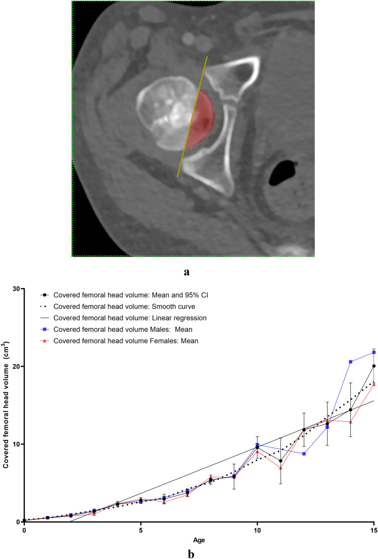
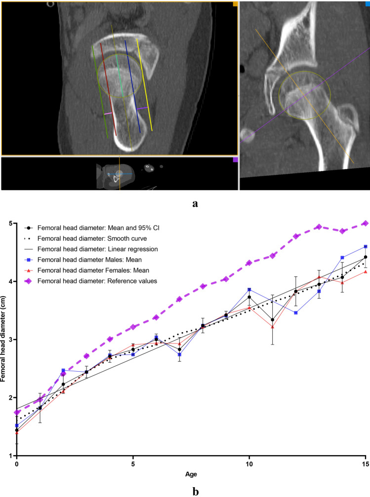
Morphology of the proximal femur was retrospectively assessed in 170 hips (85 children, < 15 years). Measurements included covered femoral head volume (CFHV), femoral head diameter (FHD), femoral head extrusion index (FHEI), coronal alpha angle (CAA), lateral centre-edge angle (LCEA), anterior (AOS) and posterior head-neck offset (POS) and femoral neck-shaft angle (FNSA). Correlation analyses as well as inter- and intra-rater reliability were performed.

RESULTS

CFHV, LCEA, FHD and AOS/POS increased with age and FHEI, CAA, and FNSA decreased with age. None of the measurements correlated with the side. AOS showed a poor correlation with gender. Rapid growth phases were observed at the age of 1, 7 and 11. The inter- and intra-rater reliability was high (range ICC 0.8-0.99 Cronbach alpha 0.86-0.99).

CONCLUSION

This data delivers a description of growth phases as well as gender and age-correlated reference values of the proximal femoral morphology that could be used by paediatricians and orthopaedic/paediatric surgeons to early diagnose proximal femur deformities and provide guidance in the planning of possible operations.

摘要

目的

在区分正常解剖变异与病理性畸形时,定义儿童/青少年每个年龄段股骨近端和股骨头的正常人体测量范围是必要的。本研究旨在根据正常无症状儿童/青少年的 3D-CT 测量值定义一组正常人体测量参数,并分析因年龄、性别和/或侧别而产生的变化。

方法

回顾性评估了 170 髋(85 名儿童,年龄<15 岁)的股骨近端形态。测量包括股骨头覆盖体积(CFHV)、股骨头直径(FHD)、股骨头挤出指数(FHEI)、冠状面 alpha 角(CAA)、外侧中心边缘角(LCEA)、前(AOS)和后股骨头颈偏移(POS)以及股骨颈干角(FNSA)。进行了相关性分析以及组内和组间可靠性分析。

结果

CFHV、LCEA、FHD 和 AOS/POS 随年龄增加而增加,而 FHEI、CAA 和 FNSA 随年龄增加而减少。任何测量均与侧别无关。AOS 与性别相关性较差。在 1 岁、7 岁和 11 岁时观察到快速生长阶段。组内和组间可靠性均较高(ICC 范围 0.8-0.99,Cronbach alpha 0.86-0.99)。

结论

本数据提供了股骨近端形态的生长阶段以及与性别和年龄相关的参考值描述,儿科医生和矫形/儿科外科医生可以使用这些数据来早期诊断股骨近端畸形,并为可能的手术计划提供指导。

https://cdn.ncbi.nlm.nih.gov/pmc/blobs/298a/8536629/1d783c4764bf/276_2021_2841_Fig6_HTML.jpg
https://cdn.ncbi.nlm.nih.gov/pmc/blobs/298a/8536629/cad68d7f7fe1/276_2021_2841_Fig1_HTML.jpg
https://cdn.ncbi.nlm.nih.gov/pmc/blobs/298a/8536629/f5c1c04e2262/276_2021_2841_Fig2_HTML.jpg
https://cdn.ncbi.nlm.nih.gov/pmc/blobs/298a/8536629/e3850235c467/276_2021_2841_Fig3_HTML.jpg
https://cdn.ncbi.nlm.nih.gov/pmc/blobs/298a/8536629/5caf29d0db1d/276_2021_2841_Fig4_HTML.jpg
https://cdn.ncbi.nlm.nih.gov/pmc/blobs/298a/8536629/04dfee380cf2/276_2021_2841_Fig5a_HTML.jpg
https://cdn.ncbi.nlm.nih.gov/pmc/blobs/298a/8536629/1d783c4764bf/276_2021_2841_Fig6_HTML.jpg

相似文献

1
Anthropometry of the proximal femur and femoral head in children/adolescents using three-dimensional computed tomography-based measurements.基于三维 CT 测量的儿童/青少年股骨近端和股骨头的人体测量学研究。
Surg Radiol Anat. 2021 Dec;43(12):2009-2023. doi: 10.1007/s00276-021-02841-3. Epub 2021 Oct 1.
2
Femoral Morphology in the Dysplastic Hip: Three-dimensional Characterizations With CT.发育性髋关节异常中的股骨形态学:CT三维特征分析
Clin Orthop Relat Res. 2017 Apr;475(4):1045-1054. doi: 10.1007/s11999-016-5119-2.
3
Anthropometric measurements of the pediatric hip using CT-based simulated anteroposterior radiographs of the pelvis.基于 CT 的骨盆前后位模拟 X 线片对小儿髋关节进行人体测量。
J Pediatr Orthop B. 2022 Jul 1;31(4):334-343. doi: 10.1097/BPB.0000000000000940. Epub 2021 Dec 2.
4
Which Two-dimensional Radiographic Measurements of Cam Femoroacetabular Impingement Best Describe the Three-dimensional Shape of the Proximal Femur?Cam 型股骨髋臼撞击症的二维放射学测量中,哪些指标最能描述股骨近端的三维形态?
Clin Orthop Relat Res. 2019 Jan;477(1):242-253. doi: 10.1097/CORR.0000000000000462.
5
Three-dimensional computerized tomographic analysis of the deformity of lateral growth disturbance of proximal femoral physis.股骨近端骨骺外侧生长紊乱畸形的三维计算机断层扫描分析
J Pediatr Orthop. 2009 Sep;29(6):540-6. doi: 10.1097/BPO.0b013e3181b2f73e.
6
Anthropometric three-dimensional computed tomography reconstruction measurements of the acetabulum in children/adolescents.儿童/青少年髋臼的人体测量三维计算机断层扫描重建测量。
Acta Orthop Belg. 2022 Mar;88(1):1-9. doi: 10.52628/88.1.01.
7
Morphologic Features of the Contralateral Femur in Patients With Unilateral Slipped Capital Femoral Epiphysis Resembles Mild Slip Deformity: A Matched Cohort Study.单侧股骨头骨骺滑脱患者对侧股骨的形态特征类似于轻度滑脱畸形:一项配对队列研究。
Clin Orthop Relat Res. 2018 Apr;476(4):890-899. doi: 10.1007/s11999.0000000000000127.
8
Reference values for proximal femoral anatomy in adolescents based on sex, physis, and imaging plane.青少年股骨近端解剖学的参考值基于性别、骨骺和成像平面。
Am J Sports Med. 2013 Sep;41(9):2074-82. doi: 10.1177/0363546513495346. Epub 2013 Jul 12.
9
Proximal femoral morphology after transtrochanteric posterior rotational osteotomy for osteonecrosis of the femoral head: A three-dimensional simulation study.转子间后旋截骨术治疗股骨头坏死术后股骨近端形态的三维模拟研究。
Orthop Traumatol Surg Res. 2020 Dec;106(8):1569-1574. doi: 10.1016/j.otsr.2020.07.003. Epub 2020 Nov 3.
10
Proximal femoral radiographic changes after lateral transtrochanteric intramedullary nail placement in children.儿童经大转子外侧髓内钉置入术后股骨近端的影像学变化
J Bone Joint Surg Am. 2003 Jul;85(7):1295-301. doi: 10.2106/00004623-200307000-00016.

引用本文的文献

1
AI-Assisted 3D Planning of CT Parameters for Personalized Femoral Prosthesis Selection in Total Hip Arthroplasty.全髋关节置换术中用于个性化股骨假体选择的CT参数的人工智能辅助3D规划
Ther Clin Risk Manag. 2025 Jun 18;21:905-916. doi: 10.2147/TCRM.S521755. eCollection 2025.
2
Management of pediatric femoral neck fractures from classification to surgery: a review of indications based on anatomic and radiographic features of the proximal femur.小儿股骨颈骨折的管理:从分类到手术——基于股骨近端解剖和影像学特征的适应证综述
EFORT Open Rev. 2025 Mar 3;10(3):125-140. doi: 10.1530/EOR-2024-0129.

本文引用的文献

1
MRI and sonography in Legg-Calvé-Perthes disease: clinical relevance of containment and influence on treatment.Legg-Calvé-Perthes病的磁共振成像与超声检查:包容的临床相关性及其对治疗的影响
J Child Orthop. 2018 Oct 1;12(5):472-479. doi: 10.1302/1863-2548.12.180033.
2
Acetabular Retroversion and Decreased Posterior Coverage Are Associated With Sports-related Posterior Hip Dislocation in Adolescents.髋臼后倾和后覆盖减少与青少年运动相关性髋关节后脱位有关。
Clin Orthop Relat Res. 2019 May;477(5):1101-1108. doi: 10.1097/CORR.0000000000000514.
3
The natural alpha angle of the femoral head-neck junction: a cross-sectional CT study in 1312 femurs.
股骨头颈交界区的自然 alpha 角:1312 例股骨的横断面 CT 研究。
Bone Joint J. 2018 May 1;100-B(5):570-578. doi: 10.1302/0301-620X.100B5.BJJ-2017-0249.R3.
4
The Anteroposterior Pelvic Radiograph: Acetabular and Femoral Measurements and Relation to Hip Pathologies.骨盆前后位X线片:髋臼和股骨测量及其与髋关节病变的关系
J Bone Joint Surg Am. 2018 Jan 3;100(1):76-85. doi: 10.2106/JBJS.17.00500.
5
Acetabular overcoverage in the horizontal plane: an underdiagnosed trigger of early hip arthritis. A CT scan study in young adults.髋臼在水平面的过度覆盖:早期髋关节炎的一个未被充分诊断的触发因素。一项针对年轻人的CT扫描研究。
Arch Orthop Trauma Surg. 2018 Jan;138(1):73-82. doi: 10.1007/s00402-017-2811-y. Epub 2017 Oct 30.
6
Vital Signs: Prevalence of Doctor-Diagnosed Arthritis and Arthritis-Attributable Activity Limitation - United States, 2013-2015.生命体征:美国2013 - 2015年医生诊断的关节炎患病率及关节炎所致活动受限情况
MMWR Morb Mortal Wkly Rep. 2017 Mar 10;66(9):246-253. doi: 10.15585/mmwr.mm6609e1.
7
Multiplanar evaluation of radiological findings associated with acetabular dysplasia and investigation of its prevalence in an Asian population: a CT-based study.髋臼发育不良相关放射学表现的多平面评估及其在亚洲人群中的患病率调查:一项基于CT的研究
BMC Musculoskelet Disord. 2017 Jan 31;18(1):50. doi: 10.1186/s12891-017-1426-3.
8
Normal radiological unossified hip joint space and femoral head size development during growth in 675 children and adolescents.675名儿童和青少年生长过程中正常的放射学未骨化髋关节间隙及股骨头大小发育情况。
Clin Anat. 2017 Mar;30(2):267-275. doi: 10.1002/ca.22807. Epub 2016 Nov 27.
9
MR Imaging with Metal-suppression Sequences for Evaluation of Total Joint Arthroplasty.使用金属抑制序列的磁共振成像用于评估全关节置换术
Radiographics. 2016 Jan-Feb;36(1):209-25. doi: 10.1148/rg.2016150075. Epub 2015 Nov 20.
10
The femoral neck-shaft angle on plain radiographs: a systematic review.X线平片上的股骨颈干角:一项系统评价
Skeletal Radiol. 2016 Jan;45(1):19-28. doi: 10.1007/s00256-015-2236-z. Epub 2015 Aug 25.