• 文献检索
  • 文档翻译
  • 深度研究
  • 学术资讯
  • Suppr Zotero 插件Zotero 插件
  • 邀请有礼
  • 套餐&价格
  • 历史记录
应用&插件
Suppr Zotero 插件Zotero 插件浏览器插件Mac 客户端Windows 客户端微信小程序
定价
高级版会员购买积分包购买API积分包
服务
文献检索文档翻译深度研究API 文档MCP 服务
关于我们
关于 Suppr公司介绍联系我们用户协议隐私条款
关注我们

Suppr 超能文献

核心技术专利:CN118964589B侵权必究
粤ICP备2023148730 号-1Suppr @ 2026

文献检索

告别复杂PubMed语法,用中文像聊天一样搜索,搜遍4000万医学文献。AI智能推荐,让科研检索更轻松。

立即免费搜索

文件翻译

保留排版,准确专业,支持PDF/Word/PPT等文件格式,支持 12+语言互译。

免费翻译文档

深度研究

AI帮你快速写综述,25分钟生成高质量综述,智能提取关键信息,辅助科研写作。

立即免费体验

钙信号在斑马鱼中脑-后脑边界形成过程中驱动细胞形态变化。

Calcium signals drive cell shape changes during zebrafish midbrain-hindbrain boundary formation.

作者信息

Sahu Srishti U, Visetsouk Mike R, Garde Ryan J, Hennes Leah, Kwas Constance, Gutzman Jennifer H

机构信息

Department of Biological Sciences, University of Wisconsin-Milwaukee, Milwaukee, WI 53201.

Department of Biological Sciences, University of Wisconsin-Milwaukee, Milwaukee, WI 53201

出版信息

Mol Biol Cell. 2017 Apr 1;28(7):875-882. doi: 10.1091/mbc.E16-08-0561. Epub 2017 Feb 1.

DOI:10.1091/mbc.E16-08-0561
PMID:28148652
原文链接:https://pmc.ncbi.nlm.nih.gov/articles/PMC5385936/
Abstract

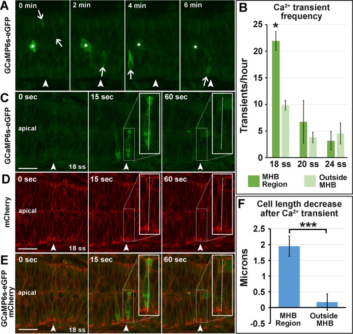
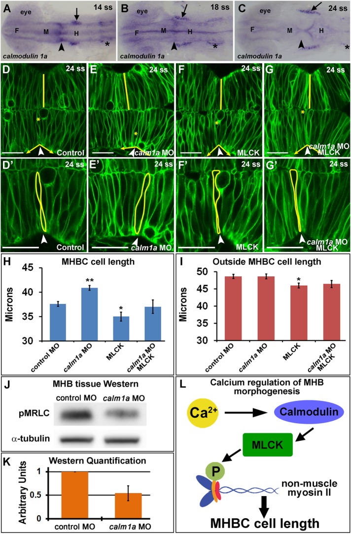
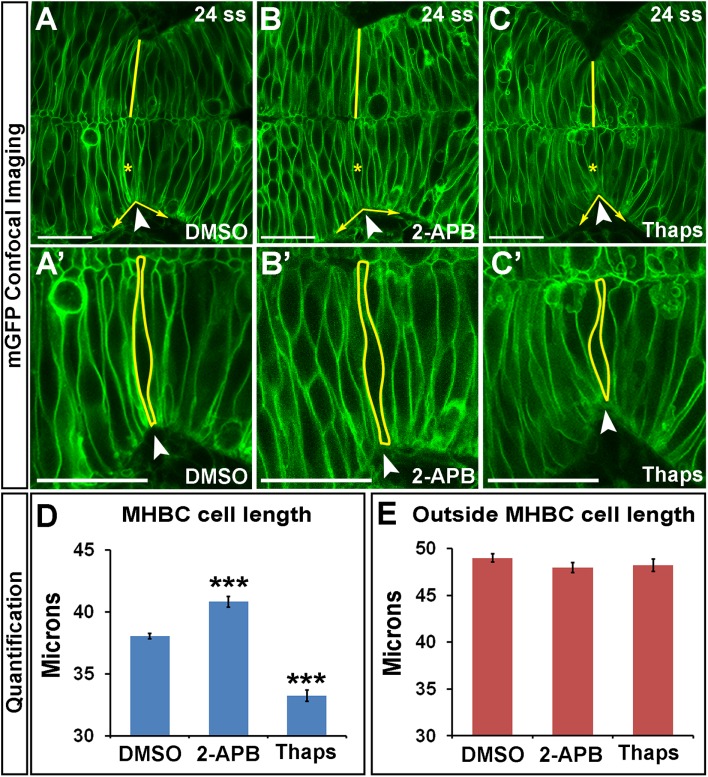
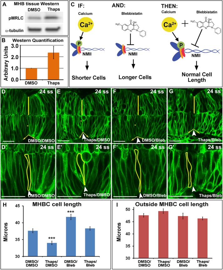
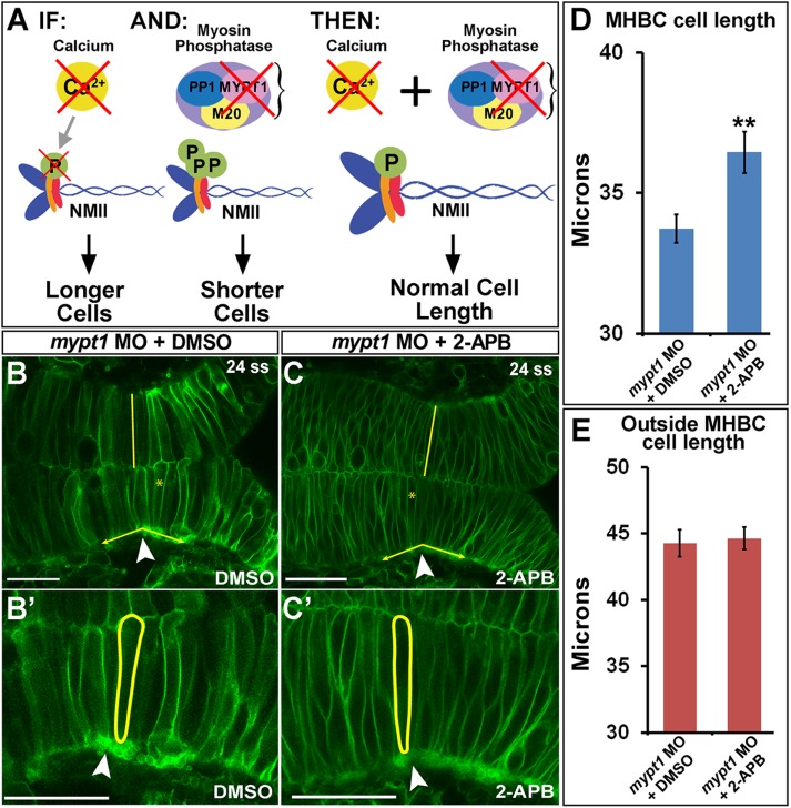
One of the first morphogenetic events in the vertebrate brain is the formation of the highly conserved midbrain-hindbrain boundary (MHB). Specific cell shape changes occur at the point of deepest constriction of the MHB, the midbrain-hindbrain boundary constriction (MHBC), and are critical for proper MHB formation. These cell shape changes are controlled by nonmuscle myosin II (NMII) motor proteins, which are tightly regulated via the phosphorylation of their associated myosin regulatory light chains (MRLCs). However, the upstream signaling pathways that initiate the regulation of NMII to mediate cell shape changes during MHB morphogenesis are not known. We show that intracellular calcium signals are critical for the regulation of cell shortening during initial MHB formation. We demonstrate that the MHB region is poised to respond to calcium transients that occur in the MHB at the onset of MHB morphogenesis and that calcium mediates phosphorylation of MRLC specifically in MHB tissue. Our results indicate that (), expressed specifically in the MHB, and myosin light chain kinase together mediate MHBC cell length. Our data suggest that modulation of NMII activity by calcium is critical for proper regulation of cell length to determine embryonic brain shape during development.

摘要

脊椎动物大脑中最早的形态发生事件之一是高度保守的中脑-后脑边界(MHB)的形成。在MHB最深收缩点,即中脑-后脑边界收缩(MHBC)处会发生特定的细胞形态变化,这些变化对于MHB的正常形成至关重要。这些细胞形态变化由非肌肉肌球蛋白II(NMII)运动蛋白控制,该蛋白通过其相关的肌球蛋白调节轻链(MRLC)的磷酸化进行严格调控。然而,在MHB形态发生过程中启动对NMII的调节以介导细胞形态变化的上游信号通路尚不清楚。我们发现细胞内钙信号对于初始MHB形成过程中细胞缩短的调节至关重要。我们证明MHB区域准备好对MHB形态发生开始时在MHB发生的钙瞬变做出反应,并且钙特异性地介导MHB组织中MRLC的磷酸化。我们的结果表明,在MHB中特异性表达的()与肌球蛋白轻链激酶共同介导MHBC细胞长度。我们的数据表明,钙对NMII活性的调节对于在发育过程中正确调节细胞长度以确定胚胎脑形态至关重要。

https://cdn.ncbi.nlm.nih.gov/pmc/blobs/95f3/5385936/3823d652370d/875fig5.jpg
https://cdn.ncbi.nlm.nih.gov/pmc/blobs/95f3/5385936/a45e5c6bc7c7/875fig1.jpg
https://cdn.ncbi.nlm.nih.gov/pmc/blobs/95f3/5385936/ca96adfc78c2/875fig2.jpg
https://cdn.ncbi.nlm.nih.gov/pmc/blobs/95f3/5385936/275fc402003e/875fig3.jpg
https://cdn.ncbi.nlm.nih.gov/pmc/blobs/95f3/5385936/a0ea474b7bd4/875fig4.jpg
https://cdn.ncbi.nlm.nih.gov/pmc/blobs/95f3/5385936/3823d652370d/875fig5.jpg

相似文献

1
Calcium signals drive cell shape changes during zebrafish midbrain-hindbrain boundary formation.钙信号在斑马鱼中脑-后脑边界形成过程中驱动细胞形态变化。
Mol Biol Cell. 2017 Apr 1;28(7):875-882. doi: 10.1091/mbc.E16-08-0561. Epub 2017 Feb 1.
2
Non-muscle myosin IIA and IIB differentially regulate cell shape changes during zebrafish brain morphogenesis.非肌肉肌球蛋白IIA和IIB在斑马鱼脑形态发生过程中对细胞形状变化发挥不同的调节作用。
Dev Biol. 2015 Jan 1;397(1):103-15. doi: 10.1016/j.ydbio.2014.10.017. Epub 2014 Oct 31.
3
Zebrafish gbx1 refines the midbrain-hindbrain boundary border and mediates the Wnt8 posteriorization signal.斑马鱼gbx1基因优化中脑-后脑边界,并介导Wnt8后化信号。
Neural Dev. 2009 Apr 2;4:12. doi: 10.1186/1749-8104-4-12.
4
Midbrain-hindbrain boundary patterning and morphogenesis are regulated by diverse grainy head-like 2-dependent pathways.中脑-后脑边界的模式形成和形态发生受多种 Grainy head-like 2 依赖性途径的调节。
Development. 2012 Feb;139(3):525-36. doi: 10.1242/dev.066522.
5
Gbx2 functions as a transcriptional repressor to regulate the specification and morphogenesis of the mid-hindbrain junction in a dosage- and stage-dependent manner.Gbx2 作为转录抑制因子,以剂量和时间依赖的方式发挥作用,调节中后脑交界处的特化和形态发生。
Mech Dev. 2013 Nov-Dec;130(11-12):532-52. doi: 10.1016/j.mod.2013.07.004. Epub 2013 Aug 7.
6
Formation of the zebrafish midbrain-hindbrain boundary constriction requires laminin-dependent basal constriction.斑马鱼中脑-后脑边界收缩的形成需要层粘连蛋白依赖性基底收缩。
Mech Dev. 2008 Nov-Dec;125(11-12):974-83. doi: 10.1016/j.mod.2008.07.004. Epub 2008 Jul 18.
7
Knockdown of epigenetic transcriptional co-regulator Brd2a disrupts apoptosis and proper formation of hindbrain and midbrain-hindbrain boundary (MHB) region in zebrafish.敲低表观遗传转录共调节因子Brd2a会破坏斑马鱼的细胞凋亡以及后脑和中脑-后脑边界(MHB)区域的正常形成。
Mech Dev. 2017 Aug;146:10-30. doi: 10.1016/j.mod.2017.05.003. Epub 2017 May 23.
8
Novel mechanisms that pattern and shape the midbrain-hindbrain boundary.中脑-后脑边界的新型模式形成和塑造机制。
Cell Mol Life Sci. 2013 Sep;70(18):3365-74. doi: 10.1007/s00018-012-1240-x. Epub 2013 Jan 10.
9
Combinatorial Wnt control of zebrafish midbrain-hindbrain boundary formation.Wnt组合调控斑马鱼中脑-后脑边界的形成。
Mech Dev. 2004 May;121(5):437-47. doi: 10.1016/j.mod.2004.03.026.
10
MicroRNA-9 directs late organizer activity of the midbrain-hindbrain boundary.微小RNA-9指导中脑-后脑边界的晚期组织者活性。
Nat Neurosci. 2008 Jun;11(6):641-8. doi: 10.1038/nn.2115. Epub 2008 May 4.

引用本文的文献

1
Calcium transients regulate the apical emergence of basally located progenitors during Xenopus skin development.在非洲爪蟾皮肤发育过程中,钙瞬变调节位于基部的祖细胞的顶端出现。
Nat Commun. 2025 Jul 19;16(1):6650. doi: 10.1038/s41467-025-61610-7.
2
The transcription factor Jun is necessary for optic nerve regeneration in larval zebrafish.转录因子Jun对斑马鱼幼体的视神经再生至关重要。
PLoS One. 2025 Mar 10;20(3):e0313534. doi: 10.1371/journal.pone.0313534. eCollection 2025.
3
myh9b is a critical non-muscle myosin II encoding gene that interacts with myh9a and myh10 during zebrafish development in both compensatory and redundant pathways.

本文引用的文献

1
Cell-Autonomous Ca(2+) Flashes Elicit Pulsed Contractions of an Apical Actin Network to Drive Apical Constriction during Neural Tube Closure.细胞自主的钙离子闪烁引发顶端肌动蛋白网络的脉冲收缩,从而在神经管闭合过程中驱动顶端收缩。
Cell Rep. 2015 Dec 15;13(10):2189-202. doi: 10.1016/j.celrep.2015.11.017. Epub 2015 Dec 7.
2
Genetic compensation induced by deleterious mutations but not gene knockdowns.有害突变而非基因敲低诱导的遗传补偿。
Nature. 2015 Aug 13;524(7564):230-3. doi: 10.1038/nature14580. Epub 2015 Jul 13.
3
Calm1 signaling pathway is essential for the migration of mouse precerebellar neurons.
myh9b是一个关键的非肌肉肌球蛋白II编码基因,在斑马鱼发育过程中,它在补偿和冗余途径中与myh9a和myh10相互作用。
G3 (Bethesda). 2025 Jan 8;15(1). doi: 10.1093/g3journal/jkae260.
4
CRISPR-RfxCas13d screening uncovers Bckdk as a post-translational regulator of the maternal-to-zygotic transition in teleosts.CRISPR-RfxCas13d筛选揭示Bckdk是硬骨鱼母源-合子转换的翻译后调节因子。
bioRxiv. 2024 May 23:2024.05.22.595167. doi: 10.1101/2024.05.22.595167.
5
Deciphering the Calcium Code: A Review of Calcium Activity Analysis Methods Employed to Identify Meaningful Activity in Early Neural Development.解读钙信号密码:用于识别早期神经发育中有意义活动的钙活性分析方法综述
Biomolecules. 2024 Jan 22;14(1):138. doi: 10.3390/biom14010138.
6
Epithelial restitution in 3D - Revealing biomechanical and physiochemical dynamics in intestinal organoids via fs laser nanosurgery.三维上皮修复——通过飞秒激光纳米手术揭示肠道类器官中的生物力学和物理化学动力学
iScience. 2023 Oct 5;26(11):108139. doi: 10.1016/j.isci.2023.108139. eCollection 2023 Nov 17.
7
Mechanosensitive calcium flashes promote sustained RhoA activation during tight junction remodeling.机械敏感性钙闪光促进紧密连接重塑过程中 RhoA 的持续激活。
J Cell Biol. 2022 Apr 4;221(4). doi: 10.1083/jcb.202105107. Epub 2022 Mar 7.
8
Identification of Zebrafish Calcium Toolkit Genes and their Expression in the Brain.鉴定斑马鱼钙工具基因及其在大脑中的表达。
Genes (Basel). 2019 Mar 18;10(3):230. doi: 10.3390/genes10030230.
9
A simple mechanochemical model for calcium signalling in embryonic epithelial cells.胚胎上皮细胞中钙信号传导的简单机械化学模型。
J Math Biol. 2019 Jun;78(7):2059-2092. doi: 10.1007/s00285-019-01333-8. Epub 2019 Mar 2.
10
Calcium Signaling in Vertebrate Development and Its Role in Disease.脊椎动物发育中的钙信号及其在疾病中的作用。
Int J Mol Sci. 2018 Oct 30;19(11):3390. doi: 10.3390/ijms19113390.
钙调蛋白1信号通路对小鼠小脑前体神经元的迁移至关重要。
Development. 2015 Jan 15;142(2):375-84. doi: 10.1242/dev.112680. Epub 2014 Dec 17.
4
Non-muscle myosin IIA and IIB differentially regulate cell shape changes during zebrafish brain morphogenesis.非肌肉肌球蛋白IIA和IIB在斑马鱼脑形态发生过程中对细胞形状变化发挥不同的调节作用。
Dev Biol. 2015 Jan 1;397(1):103-15. doi: 10.1016/j.ydbio.2014.10.017. Epub 2014 Oct 31.
5
Structural diversity of calmodulin binding to its target sites.钙调蛋白与其靶位点结合的结构多样性。
FEBS J. 2013 Nov;280(21):5551-65. doi: 10.1111/febs.12296. Epub 2013 May 13.
6
Necessary role for intracellular Ca2+ transients in initiating the apical-basolateral thinning of enveloping layer cells during the early blastula period of zebrafish development.在斑马鱼胚胎发育的早期囊胚期,细胞内 Ca2+ 瞬变在启动包裹层细胞的顶端-基底外侧变薄中发挥必要作用。
Dev Growth Differ. 2011 Jun;53(5):679-96. doi: 10.1111/j.1440-169X.2011.01275.x.
7
Tau regulates the subcellular localization of calmodulin.tau 调节钙调蛋白的亚细胞定位。
Biochem Biophys Res Commun. 2011 May 13;408(3):500-4. doi: 10.1016/j.bbrc.2011.04.082. Epub 2011 Apr 21.
8
On the origin and evolution of the tripartite brain.论三脑区的起源与演化
Brain Behav Evol. 2010;76(1):3-10. doi: 10.1159/000320218. Epub 2010 Sep 30.
9
Epithelial relaxation mediated by the myosin phosphatase regulator Mypt1 is required for brain ventricle lumen expansion and hindbrain morphogenesis.肌球蛋白磷酸酶调节因子 Mypt1 介导的上皮松弛对于脑室管腔扩张和后脑形态发生是必需的。
Development. 2010 Mar;137(5):795-804. doi: 10.1242/dev.042705.
10
Non-muscle myosin II takes centre stage in cell adhesion and migration.非肌肉肌球蛋白II在细胞黏附和迁移中起核心作用。
Nat Rev Mol Cell Biol. 2009 Nov;10(11):778-90. doi: 10.1038/nrm2786.