Mosleh Mogeeb Ahmed Ahmed, Baba Mohd Sapiyan, Malek Sorayya, Almaktari Rasheed A
Software Engineering Department, Faculty of Engineering & Information Technology, Taiz University, 6169, Taiz, Yemen.
Institute of Biological Sciences, Faculty of Science, University of Malaya, 50603, Kuala Lumpur, Malaysia.
BMC Bioinformatics. 2016 Dec 22;17(Suppl 19):499. doi: 10.1186/s12859-016-1370-5.
Cephalometric analysis and measurements of skull parameters using X-Ray images plays an important role in predicating and monitoring orthodontic treatment. Manual analysis and measurements of cephalometric is considered tedious, time consuming, and subjected to human errors. Several cephalometric systems have been developed to automate the cephalometric procedure; however, no clear insights have been reported about reliability, performance, and usability of those systems. This study utilizes some techniques to evaluate reliability, performance, and usability metric using SUS methods of the developed cephalometric system which has not been reported in previous studies.
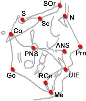
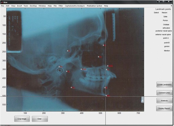
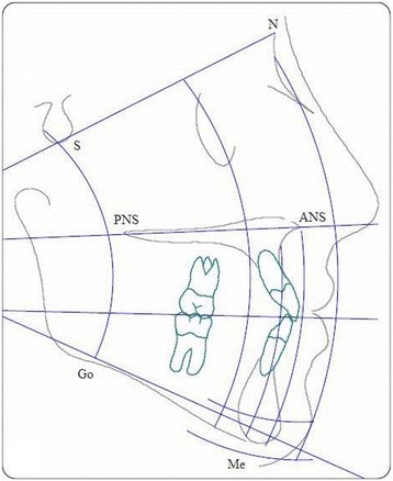
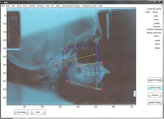
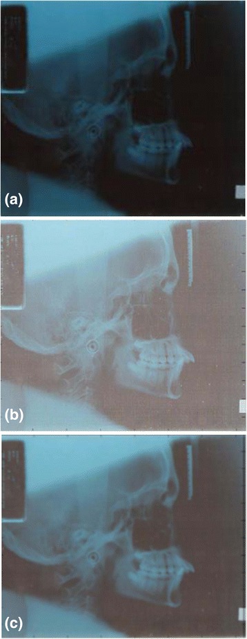
In this study a novel system named Ceph-X is developed to computerize the manual tasks of orthodontics during cephalometric measurements. Ceph-X is developed by using image processing techniques with three main models: enhancements X-ray image model, locating landmark model, and computation model. Ceph-X was then evaluated by using X-ray images of 30 subjects (male and female) obtained from University of Malaya hospital. Three orthodontics specialists were involved in the evaluation of accuracy to avoid intra examiner error, and performance for Ceph-X, and 20 orthodontics specialists were involved in the evaluation of the usability, and user satisfaction for Ceph-X by using the SUS approach.
Statistical analysis for the comparison between the manual and automatic cephalometric approaches showed that Ceph-X achieved a great accuracy approximately 96.6%, with an acceptable errors variation approximately less than 0.5 mm, and 1°. Results showed that Ceph-X increased the specialist performance, and minimized the processing time to obtain cephalometric measurements of human skull. Furthermore, SUS analysis approach showed that Ceph-X has an excellent usability user's feedback.
The Ceph-X has proved its reliability, performance, and usability to be used by orthodontists for the analysis, diagnosis, and treatment of cephalometric.
使用X射线图像进行头影测量分析和颅骨参数测量在正畸治疗的预测和监测中起着重要作用。头影测量的手动分析和测量被认为是繁琐、耗时且容易出现人为误差的。已经开发了几种头影测量系统来使头影测量程序自动化;然而,关于这些系统的可靠性、性能和可用性尚未有明确的见解报道。本研究利用一些技术,采用SUS方法评估已开发的头影测量系统的可靠性、性能和可用性指标,这在以前的研究中尚未报道。
在本研究中,开发了一种名为Ceph-X的新型系统,以实现头影测量过程中正畸手动任务的计算机化。Ceph-X是通过使用图像处理技术开发的,有三个主要模型:增强X射线图像模型、定位标志点模型和计算模型。然后使用从马来亚大学医院获得的30名受试者(男性和女性)的X射线图像对Ceph-X进行评估。三名正畸专家参与了Ceph-X准确性的评估以避免检查者内部误差和性能评估,20名正畸专家参与了通过SUS方法对Ceph-X的可用性和用户满意度的评估。
手动和自动头影测量方法之间比较的统计分析表明,Ceph-X达到了约96.6%的高精度,误差变化可接受,约小于0.5毫米和1°。结果表明,Ceph-X提高了专家的工作效率,并将获取人类颅骨的头影测量结果的处理时间降至最低。此外,SUS分析方法表明Ceph-X具有出色的可用性用户反馈。
Ceph-X已证明其可靠性、性能和可用性,可供正畸医生用于头影测量的分析、诊断和治疗。