Sassu Elena L, Ladinig Andrea, Talker Stephanie C, Stadler Maria, Knecht Christian, Stein Heiko, Frömbling Janna, Richter Barbara, Spergser Joachim, Ehling-Schulz Monika, Graage Robert, Hennig-Pauka Isabel, Gerner Wilhelm
University Clinic for Swine, Department of Farm Animals and Veterinary Public Health, University of Veterinary Medicine, Vienna, Austria.
Institute of Immunology, Department of Pathobiology, University of Veterinary Medicine Vienna, Vienna, Austria.
Vet Res. 2017 Feb 6;48(1):4. doi: 10.1186/s13567-017-0411-z.
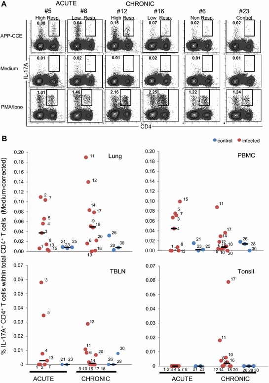
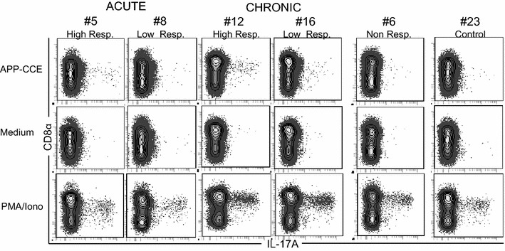
Porcine contagious pleuropneumonia caused by Actinobacillus pleuropneumoniae (APP) remains one of the major causes of poor growth performance and respiratory disease in pig herds. While the role of antibodies against APP has been intensely studied, the porcine T cell response remains poorly characterized. To address this, pigs were intranasally infected with APP serotype 2 and euthanized during the acute phase [6-10 days post-infection (dpi)] or the chronic phase of APP infection (27-31 dpi). Lymphocytes isolated from blood, tonsils, lung tissue and tracheobronchial lymph nodes were analyzed by intracellular cytokine staining (ICS) for IL-17A, IL-10 and TNF-α production after in vitro stimulation with crude capsular extract (CCE) of the APP inoculation strain. This was combined with cell surface staining for the expression of CD4, CD8α and TCR-γδ. Clinical records, microbiological investigations and pathological findings confirmed the induction of a subclinical APP infection. ICS-assays revealed the presence of APP-CCE specific CD4CD8α IL-17A-producing T cells in blood and lung tissue in most infected animals during the acute and chronic phase of infection and a minor fraction of these cells co-produced TNF-α. APP-CCE specific IL-17A-producing γδ T cells could not be found and APP-CCE specific IL-10-producing CD4 T cells were present in various organs but only in a few infected animals. The frequency of identified putative Th17 cells (CD4CD8αIL-17A) in lung and blood correlated positively with lung lesion scores and APP-specific antibody titers during the chronic phase. These results suggest a potential role of Th17 cells in the immune pathogenesis of APP infection.
由胸膜肺炎放线杆菌(APP)引起的猪传染性胸膜肺炎仍是猪群生长性能不佳和呼吸道疾病的主要原因之一。虽然针对APP抗体的作用已得到深入研究,但猪T细胞反应的特征仍不清楚。为了解决这个问题,给猪鼻内接种APP血清型2,并在急性期(感染后6 - 10天)或APP感染的慢性期(27 - 31天)实施安乐死。从血液、扁桃体、肺组织和气管支气管淋巴结分离的淋巴细胞,在体外用接种菌株的粗荚膜提取物(CCE)刺激后,通过细胞内细胞因子染色(ICS)分析IL - 17A、IL - 10和TNF -α的产生情况。这与细胞表面染色检测CD4、CD8α和TCR -γδ的表达相结合。临床记录、微生物学调查和病理结果证实了亚临床APP感染的诱导。ICS分析显示,在感染的急性期和慢性期,大多数感染动物的血液和肺组织中存在APP - CCE特异性CD4CD8α IL - 17A产生T细胞,其中一小部分细胞还共同产生TNF -α。未发现APP - CCE特异性IL - 17A产生γδ T细胞,APP - CCE特异性IL - 10产生CD4 T细胞存在于各个器官,但仅在少数感染动物中存在。在慢性期,肺和血液中鉴定出的假定Th17细胞(CD4CD8αIL - 17A)频率与肺病变评分和APP特异性抗体滴度呈正相关。这些结果表明Th17细胞在APP感染的免疫发病机制中可能起作用。