• 文献检索
  • 文档翻译
  • 深度研究
  • 学术资讯
  • Suppr Zotero 插件Zotero 插件
  • 邀请有礼
  • 套餐&价格
  • 历史记录
应用&插件
Suppr Zotero 插件Zotero 插件浏览器插件Mac 客户端Windows 客户端微信小程序
定价
高级版会员购买积分包购买API积分包
服务
文献检索文档翻译深度研究API 文档MCP 服务
关于我们
关于 Suppr公司介绍联系我们用户协议隐私条款
关注我们

Suppr 超能文献

核心技术专利:CN118964589B侵权必究
粤ICP备2023148730 号-1Suppr @ 2026

文献检索

告别复杂PubMed语法,用中文像聊天一样搜索,搜遍4000万医学文献。AI智能推荐,让科研检索更轻松。

立即免费搜索

文件翻译

保留排版,准确专业,支持PDF/Word/PPT等文件格式,支持 12+语言互译。

免费翻译文档

深度研究

AI帮你快速写综述,25分钟生成高质量综述,智能提取关键信息,辅助科研写作。

立即免费体验

内镜光学相干断层扫描血管造影微血管特征与 Barrett 食管异型增生(附视频)。

Endoscopic optical coherence tomography angiography microvascular features associated with dysplasia in Barrett's esophagus (with video).

机构信息

Department of Electrical Engineering and Computer Science and Research Laboratory of Electronics, Massachusetts Institute of Technology, Cambridge, Massachusetts, USA.

VA Boston Healthcare System, Boston, Massachusetts, USA.

出版信息

Gastrointest Endosc. 2017 Sep;86(3):476-484.e3. doi: 10.1016/j.gie.2017.01.034. Epub 2017 Feb 5.

DOI:10.1016/j.gie.2017.01.034
PMID:28167119
原文链接:https://pmc.ncbi.nlm.nih.gov/articles/PMC5545067/
Abstract

BACKGROUND AND AIMS

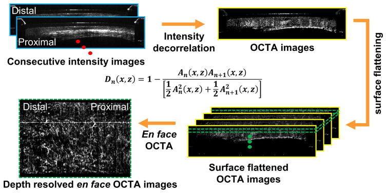
Angiogenesis is associated with neoplastic progression of Barrett's esophagus (BE). Volumetric optical coherence tomography angiography (OCTA) visualizes subsurface microvasculature without exogenous contrast agents. We investigated the association of OCTA microvascular features with low-grade dysplasia (LGD) and high-grade dysplasia (HGD).

METHODS

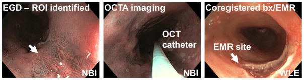
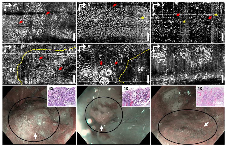
Fifty-two patients undergoing BE surveillance or endoscopic eradication therapies for dysplasia were imaged using volumetric OCTA and corresponding histologic diagnoses wre obtained to yield 97 data sets (nondysplastic BE [NDBE], 74; LGD, 10; HGD, 13). After evaluating OCTA image quality, 54 datasets (NDBE, 35; LGD, 8; HGD, 11) from 32 patients were used to develop a training and reading protocol. The association of abnormal vessel branching and heterogeneous vessel size with LGD/HGD and a regular honeycomb vessel pattern with NDBE were investigated.

RESULTS

Blinded OCTA reading of 41 OCTA datasets (NDBE, 27; LGD, 7; HGD, 7) was performed by readers with various levels of OCT/OCTA experience including 3 OCT trainees, 1 gastroenterologist, and 2 gastroenterology fellows. Among the 6 readers, OCTA features of abnormal vessel branching and heterogeneous vessel size had an overall 94% sensitivity (95% CI, 89-99) and 69% specificity (95% CI, 62-76) for differentiating LGD/HGD versus NDBE with a mean reading time of 45 seconds per data set and moderate (kappa = .58) interobserver agreement.

CONCLUSIONS

Volumetric en face OCTA imaging enables rapid examination of depth resolved microvascular features with near-microscopic resolution. OCTA can visualize microvascular features associated with LGD/HGD with high accuracy, which motivates new technologic advances and future studies investigating the diagnostic performance of OCTA.

摘要

背景和目的

血管生成与 Barrett 食管(BE)的肿瘤进展相关。容积光学相干断层扫描血管造影(OCTA)可在不使用外源性对比剂的情况下可视化皮下微血管。我们研究了 OCTA 微血管特征与低级别异型增生(LGD)和高级别异型增生(HGD)之间的关系。

方法

对 52 例接受 BE 监测或内镜下异型增生治疗的患者进行了容积 OCTA 成像,并获得了相应的组织学诊断,共获得了 97 组数据(非异型增生 BE [NDBE] 74 例;LGD 10 例;HGD 13 例)。在评估 OCTA 图像质量后,从 32 例患者中选择了 54 组(NDBE 35 组;LGD 8 组;HGD 11 组)数据来制定培训和阅读方案。研究了异常血管分支和不均匀血管大小与 LGD/HGD 的关系,以及规则的蜂窝状血管模式与 NDBE 的关系。

结果

由具有不同 OCT/OCTA 经验的 6 位读者对 41 组 OCTA 数据(NDBE 27 组;LGD 7 组;HGD 7 组)进行了盲法 OCTA 阅读,其中包括 3 位 OCT 学员、1 位胃肠病学家和 2 位胃肠病学研究员。在这 6 位读者中,异常血管分支和不均匀血管大小的 OCTA 特征在区分 LGD/HGD 与 NDBE 时的总体敏感性为 94%(95%CI,89-99),特异性为 69%(95%CI,62-76),平均阅读时间为每个数据集 45 秒,观察者间一致性中等(kappa=0.58)。

结论

容积 OCTA 成像能够快速检查深度分辨的微血管特征,具有近微观分辨率。OCTA 可以以高精度可视化与 LGD/HGD 相关的微血管特征,这激发了新技术的进步和未来研究来探索 OCTA 的诊断性能。

https://cdn.ncbi.nlm.nih.gov/pmc/blobs/d403/5545067/01e34feef550/nihms861403f3.jpg
https://cdn.ncbi.nlm.nih.gov/pmc/blobs/d403/5545067/00618fbe7ae2/nihms861403f1.jpg
https://cdn.ncbi.nlm.nih.gov/pmc/blobs/d403/5545067/a472f374c845/nihms861403f2.jpg
https://cdn.ncbi.nlm.nih.gov/pmc/blobs/d403/5545067/01e34feef550/nihms861403f3.jpg
https://cdn.ncbi.nlm.nih.gov/pmc/blobs/d403/5545067/00618fbe7ae2/nihms861403f1.jpg
https://cdn.ncbi.nlm.nih.gov/pmc/blobs/d403/5545067/a472f374c845/nihms861403f2.jpg
https://cdn.ncbi.nlm.nih.gov/pmc/blobs/d403/5545067/01e34feef550/nihms861403f3.jpg

相似文献

1
Endoscopic optical coherence tomography angiography microvascular features associated with dysplasia in Barrett's esophagus (with video).内镜光学相干断层扫描血管造影微血管特征与 Barrett 食管异型增生(附视频)。
Gastrointest Endosc. 2017 Sep;86(3):476-484.e3. doi: 10.1016/j.gie.2017.01.034. Epub 2017 Feb 5.
2
Progression from low-grade dysplasia to malignancy in patients with Barrett's esophagus diagnosed by two or more pathologists.由两名或更多病理学家诊断的巴雷特食管患者从低度发育异常进展为恶性肿瘤。
World J Gastroenterol. 2016 Oct 21;22(39):8831-8843. doi: 10.3748/wjg.v22.i39.8831.
3
Optical coherence tomography to identify intramucosal carcinoma and high-grade dysplasia in Barrett's esophagus.光学相干断层扫描用于识别巴雷特食管中的黏膜内癌和高级别异型增生。
Clin Gastroenterol Hepatol. 2006 Jan;4(1):38-43. doi: 10.1053/S1542-3565(05)00746-9.
4
Diagnosis and Management of Low-Grade Dysplasia in Barrett's Esophagus: Expert Review From the Clinical Practice Updates Committee of the American Gastroenterological Association.巴雷特食管低级别异型增生的诊断和管理:美国胃肠病学会临床实践更新委员会的专家综述。
Gastroenterology. 2016 Nov;151(5):822-835. doi: 10.1053/j.gastro.2016.09.040. Epub 2016 Oct 1.
5
Risk of malignant progression in Barrett's esophagus indefinite for dysplasia.巴雷特食管不典型增生不能确定时的恶性进展风险
Dis Esophagus. 2017 Mar 1;30(3):1-5. doi: 10.1093/dote/dow025.
6
The diagnosis of low-grade dysplasia in Barrett's esophagus and its implications for disease progression.巴雷特食管低度发育异常的诊断及其对疾病进展的影响。
Am J Gastroenterol. 2000 Dec;95(12):3383-7. doi: 10.1111/j.1572-0241.2000.03348.x.
7
Association between length of Barrett's esophagus and risk of high-grade dysplasia or adenocarcinoma in patients without dysplasia.巴雷特食管长度与无异型增生患者高级别异型增生或腺癌风险的关系。
Clin Gastroenterol Hepatol. 2013 Nov;11(11):1430-6. doi: 10.1016/j.cgh.2013.05.007. Epub 2013 May 22.
8
Barrett Esophagus Length, Nodularity, and Low-grade Dysplasia are Predictive of Progression to Esophageal Adenocarcinoma.巴雷特食管长度、结节和低级别异型增生是食管腺癌进展的预测因素。
J Clin Gastroenterol. 2019 May/Jun;53(5):361-365. doi: 10.1097/MCG.0000000000001027.
9
Assessment of Barrett's esophagus and dysplasia with ultrahigh-speed volumetric en face and cross-sectional optical coherence tomography.应用超高速度容积式内镜和横截面光学相干断层扫描技术评估 Barrett 食管及其异型增生。
Endoscopy. 2019 Apr;51(4):355-359. doi: 10.1055/a-0725-7995. Epub 2018 Sep 27.
10
Risk factors for progression of low-grade dysplasia in patients with Barrett's esophagus.巴雷特食管患者低级别上皮内瘤变进展的危险因素。
Gastroenterology. 2011 Oct;141(4):1179-86, 1186.e1. doi: 10.1053/j.gastro.2011.06.055. Epub 2011 Jun 30.

引用本文的文献

1
Optical Coherence Tomography Angiography of the Intestine: How to Prevent Motion Artifacts in Open and Laparoscopic Surgery?肠道光学相干断层扫描血管造影:如何在开放手术和腹腔镜手术中预防运动伪影?
Life (Basel). 2023 Mar 6;13(3):705. doi: 10.3390/life13030705.
2
En-face polarization-sensitive optical coherence tomography to characterize early-stage esophageal cancer and determine tumor margin.采用表面偏振敏感光学相干断层扫描技术对早期食管癌进行特征分析并确定肿瘤边界。
Biomed Opt Express. 2022 Aug 15;13(9):4773-4786. doi: 10.1364/BOE.463451. eCollection 2022 Sep 1.
3
Research progress on the application of optical coherence tomography in the field of oncology.

本文引用的文献

1
Identification of volumetric laser endomicroscopy features predictive for early neoplasia in Barrett's esophagus using high-quality histological correlation.利用高质量组织学相关性鉴定预测巴雷特食管早期肿瘤形成的容积激光内镜特征。
Gastrointest Endosc. 2017 May;85(5):918-926.e7. doi: 10.1016/j.gie.2016.09.012. Epub 2016 Sep 19.
2
Circumferential optical coherence tomography angiography imaging of the swine esophagus using a micromotor balloon catheter.使用微电机球囊导管对猪食管进行圆周光学相干断层扫描血管造影成像。
Biomed Opt Express. 2016 Jul 5;7(8):2927-42. doi: 10.1364/BOE.7.002927. eCollection 2016 Aug 1.
3
光学相干断层扫描在肿瘤学领域应用的研究进展
Front Oncol. 2022 Jul 25;12:953934. doi: 10.3389/fonc.2022.953934. eCollection 2022.
4
Endoscopic optical coherence tomography angiography using inverse SNR-amplitude decorrelation features and electrothermal micro-electro-mechanical system raster scan.利用逆信噪比 - 幅度去相关特征和电热微机电系统光栅扫描的内镜光学相干断层扫描血管造影术。
Quant Imaging Med Surg. 2022 Jun;12(6):3078-3091. doi: 10.21037/qims-21-1056.
5
Quantitative analysis of vascular changes during photoimmunotherapy using speckle variance optical coherence tomography (SV-OCT).使用散斑方差光学相干断层扫描(SV-OCT)对光免疫治疗期间的血管变化进行定量分析。
Biomed Opt Express. 2021 Mar 2;12(4):1804-1820. doi: 10.1364/BOE.419163. eCollection 2021 Apr 1.
6
Advanced endoscopic imaging for detecting and guiding therapy of early neoplasias of the esophagus.用于检测和指导食管早期肿瘤治疗的先进内镜成像。
Ann N Y Acad Sci. 2020 Dec;1482(1):61-76. doi: 10.1111/nyas.14523. Epub 2020 Nov 12.
7
Advances in Doppler optical coherence tomography and angiography.多普勒光学相干断层扫描与血管造影术的进展。
Transl Biophotonics. 2019 Dec;1(1-2). doi: 10.1002/tbio.201900005. Epub 2019 Nov 20.
8
Biomedical optical imaging technology and applications: From basic research toward clinical diagnosis.生物医学光学成像技术与应用:从基础研究到临床诊断
Exp Biol Med (Maywood). 2020 Feb;245(4):269-272. doi: 10.1177/1535370220909543.
9
Optical Vaginal Biopsy Using Optical Coherence Tomography.使用光学相干断层扫描的光学阴道活检
Female Pelvic Med Reconstr Surg. 2020 Feb;26(2):155-158. doi: 10.1097/SPV.0000000000000839.
10
Comparison of optical coherence tomography angiography and narrow-band imaging using a bimodal endoscope.双通道内镜的光相干断层扫描血管造影与窄带成像对比。
J Biomed Opt. 2019 Sep;25(3):1-5. doi: 10.1117/1.JBO.25.3.032003.
ASGE Technology Committee systematic review and meta-analysis assessing the ASGE Preservation and Incorporation of Valuable Endoscopic Innovations thresholds for adopting real-time imaging-assisted endoscopic targeted biopsy during endoscopic surveillance of Barrett's esophagus.
ASGE 技术委员会系统评价和荟萃分析评估了 ASGE 保留和纳入有价值的内镜创新的标准,用于在 Barrett 食管的内镜监测中采用实时成像辅助的内镜靶向活检。
Gastrointest Endosc. 2016 Apr;83(4):684-98.e7. doi: 10.1016/j.gie.2016.01.007. Epub 2016 Feb 11.
4
Development and Validation of a Classification System to Identify High-Grade Dysplasia and Esophageal Adenocarcinoma in Barrett's Esophagus Using Narrow-Band Imaging.基于窄带成像技术的 Barrett 食管高级别上皮内瘤变及食管腺癌分类系统的建立与验证
Gastroenterology. 2016 Mar;150(3):591-8. doi: 10.1053/j.gastro.2015.11.037. Epub 2015 Nov 25.
5
Comparative diagnostic performance of volumetric laser endomicroscopy and confocal laser endomicroscopy in the detection of dysplasia associated with Barrett's esophagus.容积激光内镜显微镜与共聚焦激光内镜显微镜在检测巴雷特食管相关异型增生中的诊断性能比较
Gastrointest Endosc. 2016 May;83(5):880-888.e2. doi: 10.1016/j.gie.2015.08.050. Epub 2015 Sep 3.
6
Ultrahigh speed endoscopic optical coherence tomography for gastroenterology.用于胃肠病学的超高速内镜光学相干断层扫描
Biomed Opt Express. 2014 Nov 24;5(12):4387-404. doi: 10.1364/BOE.5.004387. eCollection 2014 Dec 1.
7
Correction of rotational distortion for catheter-based en face OCT and OCT angiography.基于导管的正面光学相干断层扫描(OCT)和OCT血管造影术旋转畸变的校正
Opt Lett. 2014 Oct 15;39(20):5973-6. doi: 10.1364/OL.39.005973.
8
Endoscopic optical coherence angiography enables 3-dimensional visualization of subsurface microvasculature.内镜光学相干血管造影术能够对皮下微血管进行三维可视化。
Gastroenterology. 2014 Dec;147(6):1219-21. doi: 10.1053/j.gastro.2014.08.034. Epub 2014 Aug 27.
9
Radiofrequency ablation vs endoscopic surveillance for patients with Barrett esophagus and low-grade dysplasia: a randomized clinical trial.射频消融与内镜监测治疗 Barrett 食管伴低级别上皮内瘤变:一项随机临床试验。
JAMA. 2014 Mar 26;311(12):1209-17. doi: 10.1001/jama.2014.2511.
10
Evaluation of microvascular density in Barrett's associated neoplasia.评估巴雷特相关肿瘤中的微血管密度。
Mod Pathol. 2013 Jan;26(1):125-30. doi: 10.1038/modpathol.2012.146. Epub 2012 Aug 24.