• 文献检索
  • 文档翻译
  • 深度研究
  • 学术资讯
  • Suppr Zotero 插件Zotero 插件
  • 邀请有礼
  • 套餐&价格
  • 历史记录
应用&插件
Suppr Zotero 插件Zotero 插件浏览器插件Mac 客户端Windows 客户端微信小程序
定价
高级版会员购买积分包购买API积分包
服务
文献检索文档翻译深度研究API 文档MCP 服务
关于我们
关于 Suppr公司介绍联系我们用户协议隐私条款
关注我们

Suppr 超能文献

核心技术专利:CN118964589B侵权必究
粤ICP备2023148730 号-1Suppr @ 2026

文献检索

告别复杂PubMed语法,用中文像聊天一样搜索,搜遍4000万医学文献。AI智能推荐,让科研检索更轻松。

立即免费搜索

文件翻译

保留排版,准确专业,支持PDF/Word/PPT等文件格式,支持 12+语言互译。

免费翻译文档

深度研究

AI帮你快速写综述,25分钟生成高质量综述,智能提取关键信息,辅助科研写作。

立即免费体验

PET/MRI对头颈部癌肿瘤体积勾画的评估

Evaluation of PET/MRI for Tumor Volume Delineation for Head and Neck Cancer.

作者信息

Wang Kyle, Mullins Brandon T, Falchook Aaron D, Lian Jun, He Kelei, Shen Dinggang, Dance Michael, Lin Weili, Sills Tiffany M, Das Shiva K, Huang Benjamin Y, Chera Bhishamjit S

机构信息

Department of Radiation Oncology, University of North Carolina Hospitals , Chapel Hill, NC , USA.

State Key Laboratory for Novel Software Technology, Nanjing University , Nanjing , China.

出版信息

Front Oncol. 2017 Jan 23;7:8. doi: 10.3389/fonc.2017.00008. eCollection 2017.

DOI:10.3389/fonc.2017.00008
PMID:28168166
原文链接:https://pmc.ncbi.nlm.nih.gov/articles/PMC5253486/
Abstract

INTRODUCTION

Computed tomography (CT), combined positron emitted tomography and CT (PET/CT), and magnetic resonance imaging (MRI) are commonly used in head and neck radiation planning. Hybrid PET/MRI has garnered attention for potential added value in cancer staging and treatment planning. Herein, we compare PET/MRI vs. planning CT for head and neck cancer gross tumor volume (GTV) delineation.

MATERIAL AND METHODS

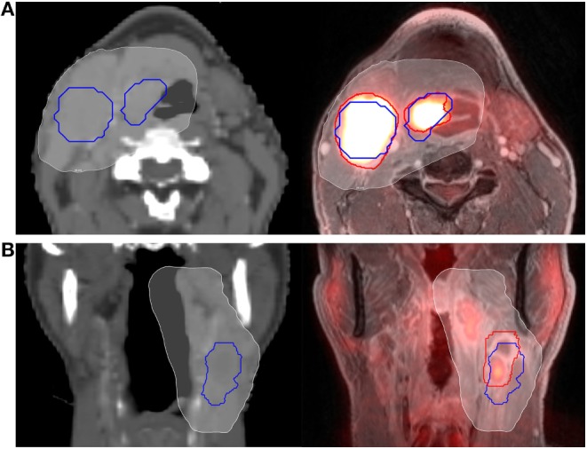
We prospectively enrolled patients with head and neck cancer treated with definitive chemoradiation to 60-70 Gy using IMRT. We performed pretreatment contrast-enhanced planning CT and gadolinium-enhanced PET/MRI. Primary and nodal volumes were delineated on planning CT (GTV-CT) prospectively before treatment and PET/MRI (GTV-PET/MRI) retrospectively after treatment. GTV-PET/MRI was compared to GTV-CT using separate rigid registrations for each tumor volume. The Dice similarity coefficient (DSC) metric evaluating spatial overlap and modified Hausdorff distance (mHD) evaluating mean orthogonal distance difference were calculated. Minimum dose to 95% of GTVs (D95) was compared.

RESULTS

Eleven patients were evaluable (10 oropharynx, 1 larynx). Nine patients had evaluable primary tumor GTVs and seven patients had evaluable nodal GTVs. Mean primary GTV-CT and GTV-PET/MRI size were 13.2 and 14.3 cc, with mean intersection 8.7 cc, DSC 0.63, and mHD 1.6 mm. D95 was 65.3 Gy for primary GTV-CT vs. 65.2 Gy for primary GTV-PET/MRI. Mean nodal GTV-CT and GTV-PET/MRI size were 19.0 and 23.0 cc, with mean intersection 14.4 cc, DSC 0.69, and mHD 2.3 mm. D95 was 62.3 Gy for both nodal GTV-CT and GTV-PET/MRI.

CONCLUSION

In this series of patients with head and neck (primarily oropharynx) cancer, PET/MRI and CT-GTVs had similar volumes (though there were individual cases with larger differences) with overall small discrepancies in spatial overlap, small mean orthogonal distance differences, and similar radiation doses.

摘要

引言

计算机断层扫描(CT)、正电子发射断层扫描与CT联合(PET/CT)以及磁共振成像(MRI)常用于头颈部放射治疗计划。混合PET/MRI在癌症分期和治疗计划中的潜在附加价值已受到关注。在此,我们比较PET/MRI与计划CT对头颈部癌大体肿瘤体积(GTV)的勾画。

材料与方法

我们前瞻性纳入接受调强放疗(IMRT)、确定性放化疗、剂量为60 - 70 Gy的头颈部癌患者。我们在治疗前进行了增强对比计划CT和钆增强PET/MRI检查。在治疗前前瞻性地在计划CT(GTV-CT)上勾画原发灶和淋巴结体积,在治疗后回顾性地在PET/MRI(GTV-PET/MRI)上勾画。对每个肿瘤体积使用单独的刚性配准将GTV-PET/MRI与GTV-CT进行比较。计算评估空间重叠的Dice相似系数(DSC)指标和评估平均正交距离差异的改良豪斯多夫距离(mHD)。比较GTVs的95%所接受的最小剂量(D95)。

结果

11例患者可评估(10例口咽癌,1例喉癌)。9例患者有可评估的原发肿瘤GTV,7例患者有可评估的淋巴结GTV。原发灶GTV-CT和GTV-PET/MRI的平均大小分别为13.2 cc和14.3 cc,平均交集为8.7 cc,DSC为0.63,mHD为1.6 mm。原发灶GTV-CT的D95为65.3 Gy,原发灶GTV-PET/MRI的D95为65.2 Gy。淋巴结GTV-CT和GTV-PET/MRI的平均大小分别为19.0 cc和23.0 cc,平均交集为14.4 cc,DSC为0.69,mHD为2.3 mm。淋巴结GTV-CT和GTV-PET/MRI的D95均为62.3 Gy。

结论

在这组头颈部(主要是口咽)癌患者中,PET/MRI和CT-GTV的体积相似(尽管个别病例差异较大),总体空间重叠差异小,平均正交距离差异小,辐射剂量相似。

https://cdn.ncbi.nlm.nih.gov/pmc/blobs/83ba/5253486/023d3a8b0cbd/fonc-07-00008-g002.jpg
https://cdn.ncbi.nlm.nih.gov/pmc/blobs/83ba/5253486/9adc6d7f34ac/fonc-07-00008-g001.jpg
https://cdn.ncbi.nlm.nih.gov/pmc/blobs/83ba/5253486/023d3a8b0cbd/fonc-07-00008-g002.jpg
https://cdn.ncbi.nlm.nih.gov/pmc/blobs/83ba/5253486/9adc6d7f34ac/fonc-07-00008-g001.jpg
https://cdn.ncbi.nlm.nih.gov/pmc/blobs/83ba/5253486/023d3a8b0cbd/fonc-07-00008-g002.jpg

相似文献

1
Evaluation of PET/MRI for Tumor Volume Delineation for Head and Neck Cancer.PET/MRI对头颈部癌肿瘤体积勾画的评估
Front Oncol. 2017 Jan 23;7:8. doi: 10.3389/fonc.2017.00008. eCollection 2017.
2
Improving the accuracy of target volume delineation by combined use of computed tomography, magnetic resonance imaging and positron emission tomography in head and neck carcinomas.通过联合使用计算机断层扫描、磁共振成像和正电子发射断层扫描提高头颈部癌靶区勾画的准确性。
J Cancer Res Ther. 2015 Oct-Dec;11(4):746-51. doi: 10.4103/0973-1482.163679.
3
Evaluating diffusion-weighted magnetic resonance imaging for target volume delineation in head and neck radiotherapy.评估扩散加权磁共振成像在头颈部放射治疗中靶区勾画的应用
J Med Imaging Radiat Oncol. 2019 Jun;63(3):399-407. doi: 10.1111/1754-9485.12866. Epub 2019 Feb 28.
4
Target volume delineation in oropharyngeal cancer: impact of PET, MRI, and physical examination.口咽癌的靶区勾画:PET、MRI 和体格检查的影响。
Int J Radiat Oncol Biol Phys. 2012 May 1;83(1):220-7. doi: 10.1016/j.ijrobp.2011.05.060. Epub 2011 Oct 27.
5
Variability of gross tumour volume delineation: MRI and CT based tumour and lymph node delineation for lung radiotherapy.大体肿瘤体积勾画的可变性:用于肺放疗的 MRI 和 CT 肿瘤及淋巴结勾画。
Radiother Oncol. 2022 Feb;167:292-299. doi: 10.1016/j.radonc.2021.11.036. Epub 2021 Dec 8.
6
Comparison of Ga-HBED-CC PSMA-PET/CT and multiparametric MRI for gross tumour volume detection in patients with primary prostate cancer based on slice by slice comparison with histopathology.基于与组织病理学逐片比较,比较镓-羟基苄基乙二胺-N,N'-二乙酸-环糊精前列腺特异性膜抗原正电子发射断层扫描/计算机断层扫描(Ga-HBED-CC PSMA-PET/CT)和多参数磁共振成像(MRI)在原发性前列腺癌患者中检测大体肿瘤体积的情况。
Theranostics. 2017 Jan 1;7(1):228-237. doi: 10.7150/thno.16638. eCollection 2017.
7
Interobserver and intermodality variability in GTV delineation on simulation CT, FDG-PET, and MR Images of Head and Neck Cancer.头颈部癌模拟CT、FDG-PET和MR图像上大体肿瘤体积(GTV)勾画的观察者间和模态间变异性
Jacobs J Radiat Oncol. 2014 Sep;1(1):006.
8
Therapeutic optimization in head and neck radiotherapy planning: Advocacy for FDG PET-CT in treatment condition.头颈部放射治疗计划的治疗优化:提倡在治疗条件下使用 FDG PET-CT。
Bull Cancer. 2022 Dec;109(12):1262-1268. doi: 10.1016/j.bulcan.2022.07.008. Epub 2022 Oct 22.
9
PET/MRI-guided GTV delineation during radiotherapy planning in patients with squamous cell carcinoma of the tongue.在舌鳞状细胞癌患者的放射治疗计划中,使用 PET/MRI 引导的 GTV 勾画。
Strahlenther Onkol. 2019 Sep;195(9):780-791. doi: 10.1007/s00066-019-01480-3. Epub 2019 Jun 18.
10
The impact of positron emission tomography on primary tumour delineation and dosimetric outcome in intensity modulated radiotherapy of early T-stage nasopharyngeal carcinoma.正电子发射断层扫描对早期T分期鼻咽癌调强放射治疗中肿瘤原发灶勾画及剂量学结果的影响
Radiat Oncol. 2016 Aug 24;11(1):109. doi: 10.1186/s13014-016-0685-8.

引用本文的文献

1
Radiographic Response Assessments and Standardized Imaging Interpretation Criteria in Head and Neck Cancer on FDG PET/CT: A Narrative Review.头颈部癌 FDG PET/CT 的影像学反应评估及标准化影像解读标准:一项叙述性综述
Cancers (Basel). 2024 Aug 21;16(16):2900. doi: 10.3390/cancers16162900.
2
Virtual tumor mapping and margin control with 3-D planning and navigation.采用三维规划与导航技术进行虚拟肿瘤映射及边界控制。
Innov Surg Sci. 2024 Apr 25;9(1):17-24. doi: 10.1515/iss-2021-0009. eCollection 2024 Mar.
3
Role of F-FDG PET/CT in Head and Neck Squamous Cell Carcinoma: Current Evidence and Innovative Applications.

本文引用的文献

1
The Impact of Diffusion-Weighted MRI on the Definition of Gross Tumor Volume in Radiotherapy of Non-Small-Cell Lung Cancer.扩散加权磁共振成像对非小细胞肺癌放疗中大体肿瘤体积定义的影响
PLoS One. 2016 Sep 9;11(9):e0162816. doi: 10.1371/journal.pone.0162816. eCollection 2016.
2
Accurate Delineation of Glioma Infiltration by Advanced PET/MR Neuro-Imaging (FRONTIER Study): A Diagnostic Study Protocol.通过先进的PET/MR神经成像精确描绘胶质瘤浸润(前沿研究):一项诊断研究方案
Neurosurgery. 2016 Oct;79(4):535-40. doi: 10.1227/NEU.0000000000001355.
3
MRI to CT Prostate Registration for Improved Targeting in Cancer External Beam Radiotherapy.
F-FDG PET/CT在头颈部鳞状细胞癌中的作用:当前证据与创新应用
Cancers (Basel). 2024 May 16;16(10):1905. doi: 10.3390/cancers16101905.
4
Impact of MRI on target volume definition in head and neck cancer patients.磁共振成像对头颈部癌症患者靶区定义的影响。
Radiat Oncol. 2023 Sep 6;18(1):148. doi: 10.1186/s13014-023-02326-0.
5
Gross tumor volume delineation in primary prostate cancer on F-PSMA-1007 PET/MRI and Ga-PSMA-11 PET/MRI.在 F-PSMA-1007 PET/MRI 和 Ga-PSMA-11 PET/MRI 上对原发性前列腺癌进行大体肿瘤体积勾画。
Cancer Imaging. 2022 Jul 22;22(1):36. doi: 10.1186/s40644-022-00475-1.
6
Exploring the impact of metabolic imaging in head and neck cancer treatment.探讨代谢成像在头颈部癌症治疗中的影响。
Head Neck. 2022 Oct;44(10):2228-2247. doi: 10.1002/hed.27131. Epub 2022 Jul 1.
7
Magnetic Fields and Cancer: Epidemiology, Cellular Biology, and Theranostics.磁场与癌症:流行病学、细胞生物学与治疗学。
Int J Mol Sci. 2022 Jan 25;23(3):1339. doi: 10.3390/ijms23031339.
8
Evaluation of Hybrid PET/MRI for Gross Tumor Volume (GTV) Delineation in Colorectal Cancer Liver Metastases Radiotherapy.用于结直肠癌肝转移放疗中大体肿瘤体积(GTV)勾画的PET/MRI融合技术评估
Cancer Manag Res. 2021 Jul 6;13:5383-5389. doi: 10.2147/CMAR.S316969. eCollection 2021.
9
Visualising the urethra for prostate radiotherapy planning.可视化尿道用于前列腺放射治疗计划。
J Med Radiat Sci. 2021 Sep;68(3):282-288. doi: 10.1002/jmrs.485. Epub 2021 May 24.
10
PET/MRI-guided GTV delineation during radiotherapy planning in patients with squamous cell carcinoma of the tongue.在舌鳞状细胞癌患者的放射治疗计划中,使用 PET/MRI 引导的 GTV 勾画。
Strahlenther Onkol. 2019 Sep;195(9):780-791. doi: 10.1007/s00066-019-01480-3. Epub 2019 Jun 18.
用于改善癌症体外放射治疗靶向的磁共振成像到计算机断层扫描的前列腺配准
IEEE J Biomed Health Inform. 2017 Jul;21(4):1015-1026. doi: 10.1109/JBHI.2016.2581881. Epub 2016 Jun 16.
4
Positron emission tomography-computed tomography versus positron emission tomography-magnetic resonance imaging for diagnosis of oral squamous cell carcinoma: A pilot study.正电子发射断层扫描-计算机断层扫描与正电子发射断层扫描-磁共振成像在口腔鳞状细胞癌诊断中的应用:一项初步研究。
J Craniomaxillofac Surg. 2015 Dec;43(10):2129-35. doi: 10.1016/j.jcms.2015.08.030. Epub 2015 Sep 11.
5
Locoregional tumour evaluation of squamous cell carcinoma in the head and neck area: a comparison between MRI, PET/CT and integrated PET/MRI.头颈部鳞状细胞癌的局部区域肿瘤评估:MRI、PET/CT与PET/MRI一体化成像的比较
Eur J Nucl Med Mol Imaging. 2016 Jan;43(1):92-102. doi: 10.1007/s00259-015-3145-z. Epub 2015 Aug 6.
6
3T MRI evaluation of large nerve perineural spread of head and neck cancers.3T磁共振成像对头颈部癌症大神经周围扩散的评估
J Med Imaging Radiat Oncol. 2015 Oct;59(5):578-85. doi: 10.1111/1754-9485.12338. Epub 2015 Jul 14.
7
Primary tumor delineation based on (18)FDG PET for locally advanced head and neck cancer treated by chemo-radiotherapy.基于(18)FDG PET对接受放化疗的局部晚期头颈癌进行原发肿瘤勾画。
Radiother Oncol. 2015 Jul;116(1):87-93. doi: 10.1016/j.radonc.2015.06.007. Epub 2015 Jun 15.
8
Response evaluation after chemoradiotherapy for advanced nodal disease in head and neck cancer using diffusion-weighted MRI and 18F-FDG-PET-CT.使用扩散加权磁共振成像(MRI)和18F-氟代脱氧葡萄糖正电子发射断层扫描-计算机断层扫描(18F-FDG-PET-CT)对头颈部癌晚期淋巴结疾病进行放化疗后的疗效评估
Oral Oncol. 2015 May;51(5):541-7. doi: 10.1016/j.oraloncology.2015.01.017. Epub 2015 Feb 26.
9
Investigation on the role of integrated PET/MRI for target volume definition and radiotherapy planning in patients with high grade glioma.正电子发射断层显像/磁共振成像(PET/MRI)融合技术在高级别胶质瘤患者靶区定义及放射治疗计划中的作用研究
Radiother Oncol. 2014 Sep;112(3):425-9. doi: 10.1016/j.radonc.2014.09.004. Epub 2014 Oct 9.
10
Use of diffusion-weighted imaging (DWI) in PET/MRI for head and neck cancer evaluation.弥散加权成像(DWI)在头颈部癌症评估中的 PET/MRI 应用。
Eur J Nucl Med Mol Imaging. 2014 Dec;41(12):2212-21. doi: 10.1007/s00259-014-2867-7. Epub 2014 Aug 5.