• 文献检索
  • 文档翻译
  • 深度研究
  • 学术资讯
  • Suppr Zotero 插件Zotero 插件
  • 邀请有礼
  • 套餐&价格
  • 历史记录
应用&插件
Suppr Zotero 插件Zotero 插件浏览器插件Mac 客户端Windows 客户端微信小程序
定价
高级版会员购买积分包购买API积分包
服务
文献检索文档翻译深度研究API 文档MCP 服务
关于我们
关于 Suppr公司介绍联系我们用户协议隐私条款
关注我们

Suppr 超能文献

核心技术专利:CN118964589B侵权必究
粤ICP备2023148730 号-1Suppr @ 2026

文献检索

告别复杂PubMed语法,用中文像聊天一样搜索,搜遍4000万医学文献。AI智能推荐,让科研检索更轻松。

立即免费搜索

文件翻译

保留排版,准确专业,支持PDF/Word/PPT等文件格式,支持 12+语言互译。

免费翻译文档

深度研究

AI帮你快速写综述,25分钟生成高质量综述,智能提取关键信息,辅助科研写作。

立即免费体验

在舌鳞状细胞癌患者的放射治疗计划中,使用 PET/MRI 引导的 GTV 勾画。

PET/MRI-guided GTV delineation during radiotherapy planning in patients with squamous cell carcinoma of the tongue.

机构信息

Department of Radiotherapy, Comprehensive Cancer Center, Bialystok, Poland.

Department of Oncology, Department of Radiotherapy, Medical University of Bialystok, Comprehensive Cancer Center, 12 Ogrodowa St., 15-027, Bialystok, Poland.

出版信息

Strahlenther Onkol. 2019 Sep;195(9):780-791. doi: 10.1007/s00066-019-01480-3. Epub 2019 Jun 18.

DOI:10.1007/s00066-019-01480-3
PMID:31214735
原文链接:https://pmc.ncbi.nlm.nih.gov/articles/PMC6704108/
Abstract

PURPOSE

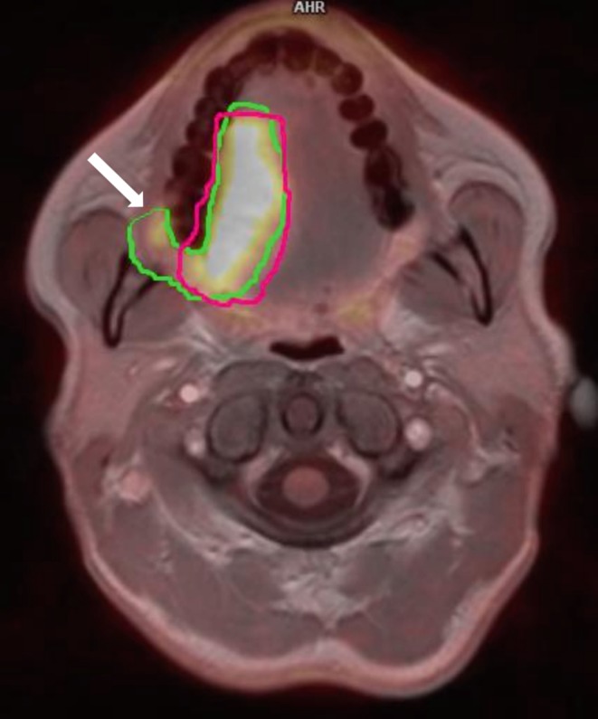
The aim of the study was to evaluate the usefulness and accuracy of 18-fluorine-labeled fluorodeoxyglucose (PET) and magnetic resonance imaging (MRI) hybrid in gross tumor volume (GTV) delineation during radiotherapy planning in patients with carcinoma of the tongue.

METHODS

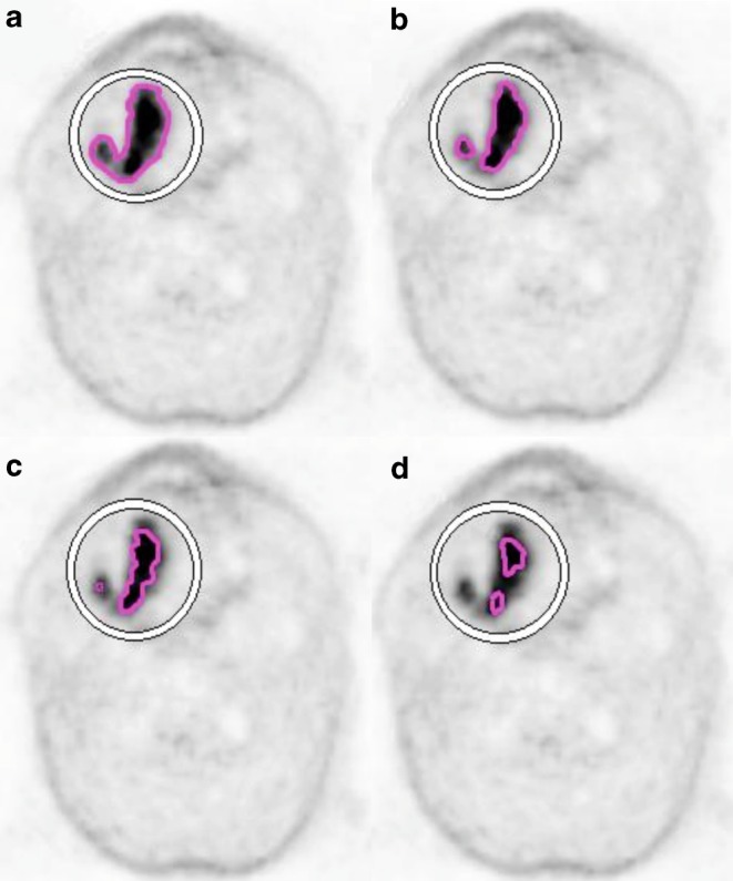
Ten patients with squamous cell carcinoma (SCC) of the tongue underwent computed tomography (CT) and PET/MRI examination. The GTV for primary tumor and lymph nodes (nGTV) were defined on CT (GTV-CT) and compared to GTVs obtained from PET (GTV-PET) and MRI (GTV-MRI) images. Two methods of GTV determination were used: visual interpretation of CT, PET (GTV-PET) and MRI images and quantitative automatic method (Syngovia, Siemens) based on a chosen threshold value (20%, 30%, 40%, 50%) of standardized uptake values (SUV) from PET examination (GTV-PET, GTV-PET, etc.). Statistical analysis of differences in GTV values obtained from CT, PET and MRI studies was performed. GTV-CT was used as a reference.

RESULTS

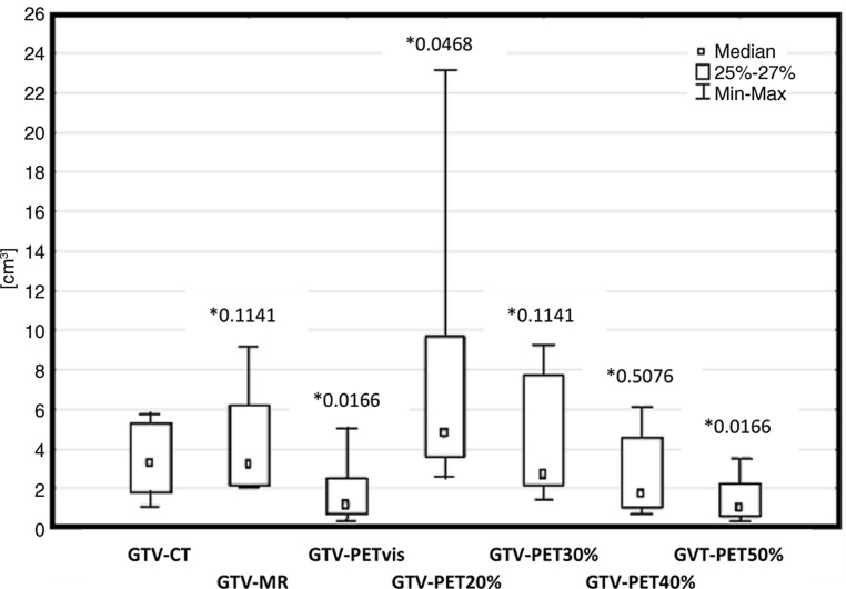
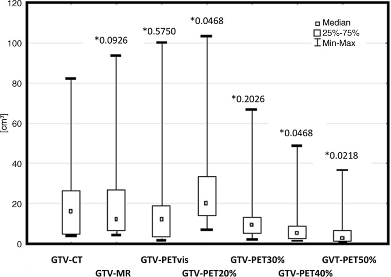
In all, 80% of GTV-MRI and 40% of GTV-PET were larger than GTV-CT. Respectively, 20% of GTV-MRI and 60% of GTV-PET were smaller than GTV-CT. Taking into account all threshold measurements, 70% of volumes were smaller than GTV-CT. GTV-PET were the most closely related volumes to GTV-CT from all threshold methods in 50% of patients. GTV-PET generated the most similar volumes in relation to GTV-CT from all PET measurements. Statistical analysis confirmed those results. Compared to nGTV-CT, 70% of nGTV-MRI and 20% of nGTV-PET were larger. The remaining nGTV-MRI and nGTV-PET measurements were smaller than nGTV-CT. Measurements of all thresholds nGTVs were smaller than nGTV-CTV in 52.5% of cases. nGTV-PET were the most closely related volumes to nGTV-CT in 40% of the cases. Statistical analysis showed that nGTV-PET (p = 0.0468), nGTV-PET (p = 0.0166), and nGTV-PET (p = 0.0166) diverge significantly from nGTV-CT results. nGTV-MRI (p = 0.1141), nGTV-PET (p = 0.2845), and nGTV-PET (p = 0.5076) were significantly related with nGTV-CT.

CONCLUSION

Combination of PET/MRI provides more information during target tumor mass delineation in radiotherapy planning of patients with SCC of the tongue than other standard imaging methods. The most frequently matching threshold value was 30% of SUV for primary tumor delineation and 30-40% of SUV for nGTV determination.

摘要

目的

本研究旨在评估在舌鳞癌患者放射治疗计划中,18 氟标记氟代脱氧葡萄糖(PET)和磁共振成像(MRI)融合在大体肿瘤体积(GTV)勾画中的有用性和准确性。

方法

10 例舌鳞癌患者行 CT 和 PET/MRI 检查。原发肿瘤和淋巴结的 GTV(nGTV)在 CT 上定义(GTV-CT),并与 PET(GTV-PET)和 MRI(GTV-MRI)图像上获得的 GTV 进行比较。使用两种 GTV 确定方法:CT、PET(GTV-PET)和 MRI 图像的视觉解释和基于所选标准摄取值(SUV)阈值(20%、30%、40%、50%)的定量自动方法(Syngovia,Siemens)(GTV-PET、GTV-PET 等)。对 CT、PET 和 MRI 研究中获得的 GTV 值进行统计学分析。以 GTV-CT 作为参考。

结果

在所有患者中,80%的 GTV-MRI 和 40%的 GTV-PET 大于 GTV-CT。相应地,20%的 GTV-MRI 和 60%的 GTV-PET 小于 GTV-CT。考虑到所有阈值测量,70%的体积小于 GTV-CT。在 50%的患者中,50%的患者中,GTV-PET 是所有阈值方法中与 GTV-CT 最相关的体积。GTV-PET 是与所有 PET 测量相关的最相似的 GTV-CT 体积。统计分析证实了这些结果。与 nGTV-CT 相比,70%的 nGTV-MRI 和 20%的 nGTV-PET 较大。其余的 nGTV-MRI 和 nGTV-PET 测量值小于 nGTV-CT。在 52.5%的病例中,所有阈值 nGTV 测量值均小于 nGTV-CTV。在 40%的病例中,nGTV-PET 是与 nGTV-CT 最相关的体积。统计分析显示,nGTV-PET(p=0.0468)、nGTV-PET(p=0.0166)和 nGTV-PET(p=0.0166)与 nGTV-CT 结果显著不同。nGTV-MRI(p=0.1141)、nGTV-PET(p=0.2845)和 nGTV-PET(p=0.5076)与 nGTV-CT 有显著相关性。

结论

在舌鳞癌患者的放射治疗计划中,PET/MRI 的联合提供了比其他标准成像方法更多的肿瘤靶区勾画信息。最常匹配的阈值是原发性肿瘤勾画的 SUV 的 30%,nGTV 确定的 SUV 的 30-40%。

https://cdn.ncbi.nlm.nih.gov/pmc/blobs/b456/6704108/188711fe0637/66_2019_1480_Fig4_HTML.jpg
https://cdn.ncbi.nlm.nih.gov/pmc/blobs/b456/6704108/ff9d04c997f7/66_2019_1480_Fig1_HTML.jpg
https://cdn.ncbi.nlm.nih.gov/pmc/blobs/b456/6704108/c5251249becc/66_2019_1480_Fig2_HTML.jpg
https://cdn.ncbi.nlm.nih.gov/pmc/blobs/b456/6704108/1bc0bc62df7f/66_2019_1480_Fig3_HTML.jpg
https://cdn.ncbi.nlm.nih.gov/pmc/blobs/b456/6704108/188711fe0637/66_2019_1480_Fig4_HTML.jpg

相似文献

1
PET/MRI-guided GTV delineation during radiotherapy planning in patients with squamous cell carcinoma of the tongue.在舌鳞状细胞癌患者的放射治疗计划中,使用 PET/MRI 引导的 GTV 勾画。
Strahlenther Onkol. 2019 Sep;195(9):780-791. doi: 10.1007/s00066-019-01480-3. Epub 2019 Jun 18.
2
Improving the accuracy of target volume delineation by combined use of computed tomography, magnetic resonance imaging and positron emission tomography in head and neck carcinomas.通过联合使用计算机断层扫描、磁共振成像和正电子发射断层扫描提高头颈部癌靶区勾画的准确性。
J Cancer Res Ther. 2015 Oct-Dec;11(4):746-51. doi: 10.4103/0973-1482.163679.
3
Tumor volume delineation in head and neck cancer with 18-fluor-fluorodeoxiglucose positron emission tomography: adaptive thresholding method applied to primary tumors and metastatic lymph nodes.头颈部癌症中 18-氟-氟代脱氧葡萄糖正电子发射断层扫描的肿瘤体积勾画:应用于原发肿瘤和转移性淋巴结的自适应阈值法。
Clin Transl Oncol. 2013 Apr;15(4):283-93. doi: 10.1007/s12094-012-0914-z. Epub 2012 Aug 3.
4
Defining PET tumor volume in cervical cancer with hybrid PET/MRI: a comparative study.利用PET/MRI混合成像定义宫颈癌的PET肿瘤体积:一项对比研究。
Nucl Med Commun. 2014 Jul;35(7):712-9. doi: 10.1097/MNM.0000000000000113.
5
The value of magnetic resonance imaging in target volume delineation of base of tongue tumours--a study using flexible surface coils.磁共振成像在舌根部肿瘤靶区勾画中的价值——使用柔性表面线圈的研究。
Radiother Oncol. 2010 Feb;94(2):161-7. doi: 10.1016/j.radonc.2009.12.021. Epub 2010 Jan 22.
6
Delineation of gross tumor volume (GTV) for radiation treatment planning of locally advanced rectal cancer using information from MRI or FDG-PET/CT: a prospective study.使用 MRI 或 FDG-PET/CT 信息对局部进展期直肠癌进行放射治疗计划的大体肿瘤体积(GTV)勾画:一项前瞻性研究。
Int J Radiat Oncol Biol Phys. 2011 Nov 15;81(4):e439-45. doi: 10.1016/j.ijrobp.2011.03.031. Epub 2011 Jun 14.
7
A multimodality segmentation framework for automatic target delineation in head and neck radiotherapy.一种用于头颈部放射治疗中自动靶区勾画的多模态分割框架。
Med Phys. 2015 Sep;42(9):5310-20. doi: 10.1118/1.4928485.
8
Intra-tumour 18F-FDG uptake heterogeneity decreases the reliability on target volume definition with positron emission tomography/computed tomography imaging.肿瘤内18F-FDG摄取异质性降低了正电子发射断层扫描/计算机断层扫描成像在靶体积定义上的可靠性。
J Med Imaging Radiat Oncol. 2015 Jun;59(3):338-45. doi: 10.1111/1754-9485.12289. Epub 2015 Feb 23.
9
PET/CT for radiotherapy treatment planning in patients with soft tissue sarcomas.正电子发射断层显像/X线计算机体层成像(PET/CT)在软组织肉瘤患者放射治疗计划中的应用
Int J Radiat Oncol Biol Phys. 2009 Nov 1;75(3):817-21. doi: 10.1016/j.ijrobp.2008.11.055. Epub 2009 Apr 20.
10
Comparison of five segmentation tools for 18F-fluoro-deoxy-glucose-positron emission tomography-based target volume definition in head and neck cancer.用于头颈部癌中基于18F-氟脱氧葡萄糖正电子发射断层扫描的靶区定义的五种分割工具的比较
Int J Radiat Oncol Biol Phys. 2007 Nov 15;69(4):1282-9. doi: 10.1016/j.ijrobp.2007.07.2333.

引用本文的文献

1
PET/MR for predicting extranodal extension of head and neck cancer.正电子发射断层显像/磁共振成像用于预测头颈癌的结外侵犯
Neuroradiology. 2025 May 21. doi: 10.1007/s00234-025-03635-9.
2
Positron Emission Tomography-Magnetic Resonance Imaging, a New Hybrid Imaging Modality for Dentomaxillofacial Malignancies-A Systematic Review.正电子发射断层扫描-磁共振成像,一种用于牙颌面恶性肿瘤的新型混合成像模式——系统评价
Diagnostics (Basel). 2025 Mar 7;15(6):654. doi: 10.3390/diagnostics15060654.
3
Role of F-FDG PET/CT in Head and Neck Squamous Cell Carcinoma: Current Evidence and Innovative Applications.

本文引用的文献

1
Upcoming imaging concepts and their impact on treatment planning and treatment response in radiation oncology.未来的影像学概念及其对放射肿瘤学治疗计划和治疗反应的影响。
Radiat Oncol. 2018 Aug 13;13(1):146. doi: 10.1186/s13014-018-1091-1.
2
Quantifying the robustness of [F]FDG-PET/CT radiomic features with respect to tumor delineation in head and neck and pancreatic cancer patients.定量评估 [F]FDG-PET/CT 放射组学特征在头颈部和胰腺癌患者肿瘤勾画中的稳健性。
Phys Med. 2018 May;49:105-111. doi: 10.1016/j.ejmp.2018.05.013. Epub 2018 May 23.
3
F-Fdg-PET-guided Planning and Re-Planning (Adaptive) Radiotherapy in Head and Neck Cancer: Current State of Art.
F-FDG PET/CT在头颈部鳞状细胞癌中的作用:当前证据与创新应用
Cancers (Basel). 2024 May 16;16(10):1905. doi: 10.3390/cancers16101905.
4
Intermodality Variability in Gross Tumor Volume Delineation for Radiation Therapy Planning in Oropharyngeal Squamous Cell Carcinoma.口咽鳞状细胞癌放射治疗计划中大体肿瘤体积勾画的模态间变异性
Adv Radiat Oncol. 2024 Feb 6;9(5):101453. doi: 10.1016/j.adro.2024.101453. eCollection 2024 May.
5
The use of PET/MRI in radiotherapy.正电子发射断层显像/磁共振成像在放射治疗中的应用。
Insights Imaging. 2024 Feb 27;15(1):63. doi: 10.1186/s13244-024-01627-6.
6
Impact of MRI on target volume definition in head and neck cancer patients.磁共振成像对头颈部癌症患者靶区定义的影响。
Radiat Oncol. 2023 Sep 6;18(1):148. doi: 10.1186/s13014-023-02326-0.
7
The Use of Radiomic Features to Predict Human Papillomavirus (HPV) Status in Head and Neck Tumors: A Review.利用影像组学特征预测头颈部肿瘤中人乳头瘤病毒(HPV)状态的综述
Cureus. 2023 Aug 31;15(8):e44476. doi: 10.7759/cureus.44476. eCollection 2023 Aug.
8
Gross tumor volume delineation in primary prostate cancer on F-PSMA-1007 PET/MRI and Ga-PSMA-11 PET/MRI.在 F-PSMA-1007 PET/MRI 和 Ga-PSMA-11 PET/MRI 上对原发性前列腺癌进行大体肿瘤体积勾画。
Cancer Imaging. 2022 Jul 22;22(1):36. doi: 10.1186/s40644-022-00475-1.
9
Exploring the impact of metabolic imaging in head and neck cancer treatment.探讨代谢成像在头颈部癌症治疗中的影响。
Head Neck. 2022 Oct;44(10):2228-2247. doi: 10.1002/hed.27131. Epub 2022 Jul 1.
10
Characterization of positioning uncertainties in PET-CT-MR trimodality solutions for radiotherapy.在用于放射治疗的 PET-CT-MR 三联体解决方案中定位不确定性的特性描述。
J Appl Clin Med Phys. 2022 Jul;23(7):e13617. doi: 10.1002/acm2.13617. Epub 2022 Apr 28.
18F-氟代脱氧葡萄糖正电子发射断层扫描引导下的头颈部癌放疗计划制定与重新计划(自适应):当前技术现状
Anticancer Res. 2017 Dec;37(12):6523-6532. doi: 10.21873/anticanres.12108.
4
Delineation of the primary tumour Clinical Target Volumes (CTV-P) in laryngeal, hypopharyngeal, oropharyngeal and oral cavity squamous cell carcinoma: AIRO, CACA, DAHANCA, EORTC, GEORCC, GORTEC, HKNPCSG, HNCIG, IAG-KHT, LPRHHT, NCIC CTG, NCRI, NRG Oncology, PHNS, SBRT, SOMERA, SRO, SSHNO, TROG consensus guidelines.喉、下咽、口咽和口腔鳞状细胞癌的原发肿瘤临床靶区(CTV-P)勾画:AIRO、CACA、DAHANCA、EORTC、GEORCC、GORTEC、HKNPCSG、HNCIG、IAG-KHT、LPRHHT、NCIC CTG、NCRI、NRG Oncology、PHNS、SBRT、SOMERA、SRO、SSHNO、TROG 共识指南。
Radiother Oncol. 2018 Jan;126(1):3-24. doi: 10.1016/j.radonc.2017.10.016. Epub 2017 Nov 24.
5
Molecular Imaging-Guided Radiotherapy for the Treatment of Head-and-Neck Squamous Cell Carcinoma: Does it Fulfill the Promises?分子影像引导放疗治疗头颈部鳞状细胞癌:是否实现了承诺?
Semin Radiat Oncol. 2018 Jan;28(1):35-45. doi: 10.1016/j.semradonc.2017.08.003.
6
Patterns-of-failure guided biological target volume definition for head and neck cancer patients: FDG-PET and dosimetric analysis of dose escalation candidate subregions.失败模式引导的头颈部癌患者生物靶区定义:FDG-PET及剂量递增候选子区域的剂量学分析
Radiother Oncol. 2017 Aug;124(2):248-255. doi: 10.1016/j.radonc.2017.07.017. Epub 2017 Jul 31.
7
Overview of the 8th Edition TNM Classification for Head and Neck Cancer.第八版头颈部癌TNM分类概述。
Curr Treat Options Oncol. 2017 Jul;18(7):40. doi: 10.1007/s11864-017-0484-y.
8
Hybrid PET/MRI-based delineation of gross tumor volume in head and neck cancer and tumor parameter analysis.基于PET/MRI融合技术对头颈部癌大体肿瘤体积的勾画及肿瘤参数分析。
Nucl Med Commun. 2017 Jul;38(7):642-649. doi: 10.1097/MNM.0000000000000687.
9
Modality-specific target definition for laryngeal and hypopharyngeal cancer on FDG-PET, CT and MRI.基于氟代脱氧葡萄糖正电子发射断层扫描(FDG-PET)、计算机断层扫描(CT)和磁共振成像(MRI)的喉癌和下咽癌的模态特异性靶区定义
Radiother Oncol. 2017 Apr;123(1):63-70. doi: 10.1016/j.radonc.2017.02.005. Epub 2017 Mar 1.
10
Evaluation of PET/MRI for Tumor Volume Delineation for Head and Neck Cancer.PET/MRI对头颈部癌肿瘤体积勾画的评估
Front Oncol. 2017 Jan 23;7:8. doi: 10.3389/fonc.2017.00008. eCollection 2017.