Ni Yawen, Teng Tao, Li Runting, Simonyi Agnes, Sun Grace Y, Lee James C
Department of Bioengineering, University of Missouri, Columbia, Missouri, United States of America.
Department of Bioengineering, University of Illinois at Chicago, Chicago, Illinois, United States of America.
PLoS One. 2017 Feb 7;12(2):e0170346. doi: 10.1371/journal.pone.0170346. eCollection 2017.
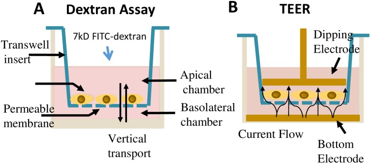
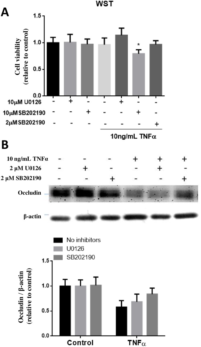
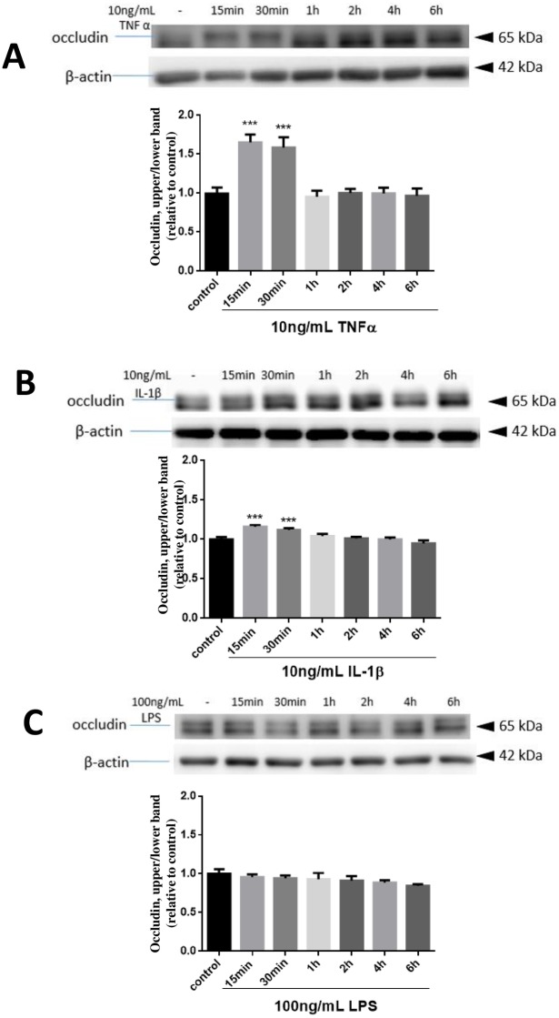
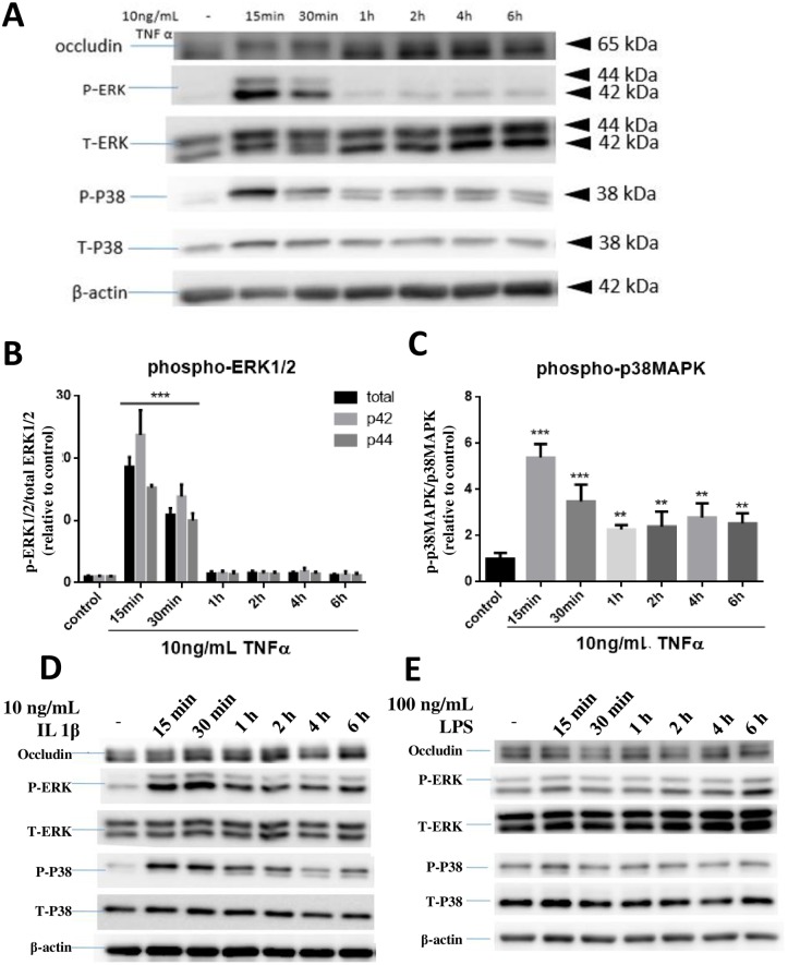
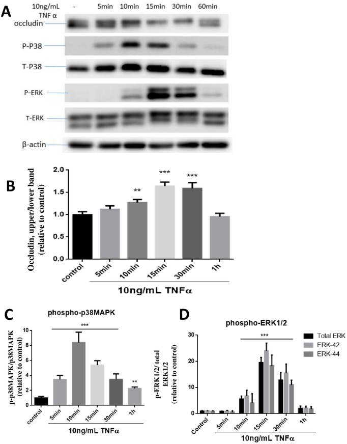
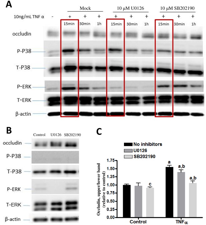
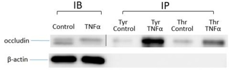
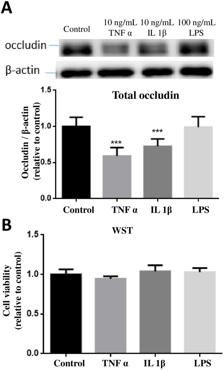
Occludin is a key tight junction (TJ) protein in cerebral endothelial cells (CECs) playing an important role in modulating blood-brain barrier (BBB) functions. This protein (65kDa) has been shown to engage in many signaling pathways and phosphorylation by both tyrosine and threonine kinases. Despite yet unknown mechanisms, pro-inflammatory cytokines and endotoxin (lipopolysaccharides, LPS) may alter TJ proteins in CECs and BBB functions. Here we demonstrate the responses of occludin in an immortalized human cerebral endothelial cell line (hCMEC/D3) to stimulation by TNFα (10 ng/mL), IL-1β (10 ng/mL) and LPS (100 ng/mL). Exposing cells to TNFα resulted in a rapid and transient upward band-shift of occludin, suggesting of an increase in phosphorylation. Exposure to IL-1β produced significantly smaller effects and LPS produced almost no effects on occludin band-shift. TNFα also caused transient stimulation of p38MAPK and ERK1/2 in hCMEC/D3 cells, and the occludin band-shift induced by TNFα was suppressed by SB202190, an inhibitor for p38MAPK, and partly by U0126, the MEK1/2-ERK1/2 inhibitor. Cells treated with TNFα and IL-1β but not LPS for 24 h resulted in a significant (p < 0.001) decrease in the expression of occludin, and the decrease could be partially blocked by SB202190, the inhibitor for p38MAPK. Treatment with TNFα also altered cell morphology and enhanced permeability of the CEC layer as measured by the FITC-dextran assay and the trans-endothelial electrical resistances (TEER). However, treatment with SB202190 alone could not effectively reverse the TNFα -induced morphology changes or the enhanced permeability changes. These results suggest that despite effects of TNFα on p38MAPK-mediated occludin phosphorylation and expression, these changes are not sufficient to avert the TNFα-induced alterations on cell morphology and permeability.
闭合蛋白是脑内皮细胞(CECs)中一种关键的紧密连接(TJ)蛋白,在调节血脑屏障(BBB)功能中发挥重要作用。这种蛋白(65kDa)已被证明参与多种信号通路,并受到酪氨酸激酶和苏氨酸激酶的磷酸化作用。尽管机制尚不清楚,但促炎细胞因子和内毒素(脂多糖,LPS)可能会改变CECs中的TJ蛋白和BBB功能。在此,我们展示了永生化人脑内皮细胞系(hCMEC/D3)中闭合蛋白对肿瘤坏死因子α(TNFα,10 ng/mL)、白细胞介素-1β(IL-1β,10 ng/mL)和LPS(100 ng/mL)刺激的反应。将细胞暴露于TNFα会导致闭合蛋白快速且短暂地上调条带迁移,提示磷酸化增加。暴露于IL-1β对闭合蛋白条带迁移的影响显著较小,而LPS对闭合蛋白条带迁移几乎没有影响。TNFα还会引起hCMEC/D3细胞中p38丝裂原活化蛋白激酶(p38MAPK)和细胞外信号调节激酶1/2(ERK1/2)的短暂刺激,并且TNFα诱导的闭合蛋白条带迁移被p38MAPK抑制剂SB202190抑制,部分被MEK1/2-ERK1/2抑制剂U0126抑制。用TNFα和IL-1β而非LPS处理细胞24小时会导致闭合蛋白表达显著(p < 0.001)降低,并且这种降低可被p38MAPK抑制剂SB202190部分阻断。用TNFα处理还会改变细胞形态,并通过异硫氰酸荧光素 - 葡聚糖测定法和跨内皮电阻(TEER)测量增强CEC层的通透性。然而,单独用SB202190处理不能有效逆转TNFα诱导的形态变化或增强的通透性变化。这些结果表明,尽管TNFα对p38MAPK介导的闭合蛋白磷酸化和表达有影响,但这些变化不足以避免TNFα诱导的细胞形态和通透性改变。