Zhao Ya-Ning, Guo Xiang-Fei, Li Jian-Min, Chen Chang-Xiang, Li Shu-Xing, Xu Cheng-Jing
Nursing and Rehabilitation College, North China University of Science and Technology, 063000, China.
The Neurosurgery of Affiliated Hospital, North China University of Science and Technology, 063000, China.
Oncotarget. 2017 Apr 4;8(14):23353-23359. doi: 10.18632/oncotarget.15058.
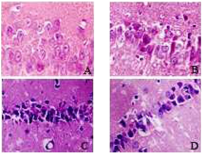
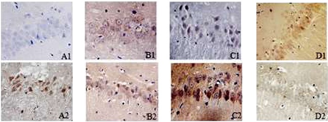
We explored the role of mTOR/autophagy pathway in the aggravation of cerebral ischemia-reperfusion nerve injury caused by intermittent hypoxia. Eighty male wistar rats were divided into four groups by the random number method: sham operation group (SO group, n=20), cerebral ischemia-reperfusion group (I/R group, n=20), intermittent hypoxia and cerebral ischemia-reperfusion group (IH+I/R group, n=20), intermittent hypoxia and cerebral ischemia-reperfusion group plus mTOR inhibitor group (inhibitor group, n=20).The results showed that compared with the SO group, HE staining showed structural damage of neurons at each time point, the immunohistochemical assay showed an increasing number of mTOR and beclin1 immune-positive cells (P<0.05) and RT-PCR showed enhanced expression of mTOR and beclin1 protein in the I/R group (P<0.05). Compared with the I/R group, HE staining showed exacerbating structural damage of neurons at each time point, the immunohistochemical assay showed an increasing number of mTOR and beclin1 immune-positive cells (P<0.05) and RT-PCR showed enhanced expression of mTOR and beclin1 protein in the IH+I/R group (P<0.05). Compared with the IH+I/R group, HE staining showed remissive structural damage of neurons at each time point, the immunohistochemical assay showed a decreasing number of mTOR immune-positive cells and a rising number of beclin1immune-positive cells (P<0.05) and RT-PCR showed weakened expression of mTOR protein and enhanced expression of beclin1 protein in the inhibitor group (P<0.05). Thence, the present study indicated that intermittent hypoxia preconditioning can aggravate the nerve injury of the global cerebral ischemia-reperfusion model, and the mechanism is associated with the activation of mTOR/autophagy pathway.
我们探讨了mTOR/自噬通路在间歇性缺氧所致脑缺血再灌注神经损伤加重中的作用。将80只雄性Wistar大鼠采用随机数字法分为四组:假手术组(SO组,n = 20)、脑缺血再灌注组(I/R组,n = 20)、间歇性缺氧合并脑缺血再灌注组(IH+I/R组,n = 20)、间歇性缺氧合并脑缺血再灌注组加mTOR抑制剂组(抑制剂组,n = 20)。结果显示,与SO组相比,HE染色显示I/R组各时间点神经元结构破坏,免疫组化检测显示mTOR和beclin1免疫阳性细胞数量增加(P<0.05),RT-PCR显示I/R组mTOR和beclin1蛋白表达增强(P<0.05)。与I/R组相比,HE染色显示IH+I/R组各时间点神经元结构破坏加重,免疫组化检测显示mTOR和beclin1免疫阳性细胞数量增加(P<0.05),RT-PCR显示IH+I/R组mTOR和beclin1蛋白表达增强(P<0.05)。与IH+I/R组相比,HE染色显示抑制剂组各时间点神经元结构破坏减轻,免疫组化检测显示mTOR免疫阳性细胞数量减少,beclin1免疫阳性细胞数量增加(P<0.05),RT-PCR显示抑制剂组mTOR蛋白表达减弱,beclin1蛋白表达增强(P<0.05)。因此,本研究表明间歇性缺氧预处理可加重全脑缺血再灌注模型的神经损伤,其机制与mTOR/自噬通路的激活有关。