Zhao Irene Shuping, Mei May Lei, Burrow Michael F, Lo Edward Chin-Man, Chu Chun-Hung
Faculty of Dentistry, The University of Hong Kong, Hong Kong 999077, China.
Melbourne Dental School, University of Melbourne, Melbourne 3010, Australia.
Int J Mol Sci. 2017 Feb 6;18(2):340. doi: 10.3390/ijms18020340.
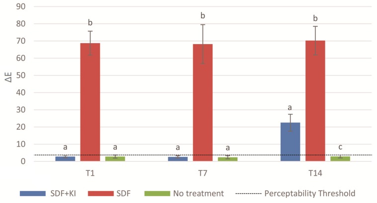
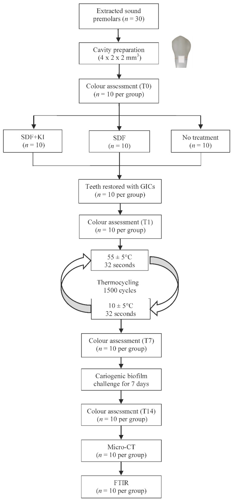
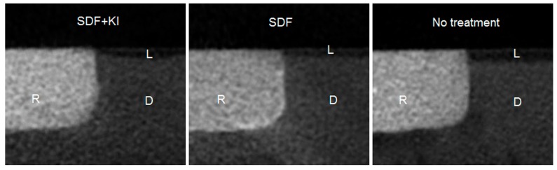
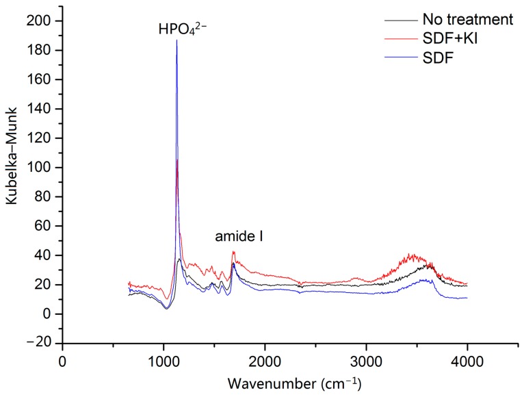
This study investigated the effect of silver diamine fluoride (SDF) and potassium iodide (KI) treatment on secondary caries prevention and tooth discolouration in glass ionomer cement (GIC) restoration. Cervical GIC restorations were done on 30 premolars with: Group 1, SDF + KI; Group 2, SDF (positive control); Group 3, no treatment (negative control). After cariogenic biofilm challenge, the demineralisation of dentine adjacent to the restoration was evaluated using micro-computed tomography (micro-CT) and Fourier transform infrared (FTIR) spectroscopy. The colour of dentine adjacent to the restoration was assessed using CIELAB system at different time points. Total colour change (∆E) was calculated and was visible if ∆E > 3.7. Micro-CT showed the outer lesion depths for Groups 1, 2 and 3 were 91 ± 7 µm, 80 ± 7 µm and 119 ± 8 µm, respectively ( < 0.001; Group 2 < Group 1 < Group 3). FTIR found that there was a significant difference in amide I-to-hydrogen phosphate ratio among the three groups ( < 0.001; Group 2 < Group 1 < Group 3). ∆E of Groups 1, 2 and 3 after biofilm challenge were 22.5 ± 4.9, 70.2 ± 8.3 and 2.9 ± 0.9, respectively ( < 0.001; Group 3 < Group 1 < Group 2). SDF + KI treatment reduced secondary caries formation on GIC restoration, but it was not as effective as SDF treatment alone. Moreover, a perceptible staining on the restoration margin was observed, but the intensity of discolouration was less than that with solely SDF treatment.
本研究调查了氟化银胺(SDF)和碘化钾(KI)处理对玻璃离子水门汀(GIC)修复体继发龋预防及牙齿变色的影响。对30颗前磨牙进行颈部GIC修复,分为:第1组,SDF+KI;第2组,SDF(阳性对照);第3组,不处理(阴性对照)。在致龋生物膜攻击后,使用微型计算机断层扫描(micro-CT)和傅里叶变换红外(FTIR)光谱评估修复体邻近牙本质的脱矿情况。在不同时间点使用CIELAB系统评估修复体邻近牙本质的颜色。计算总颜色变化(∆E),若∆E>3.7则可见。Micro-CT显示,第1、2和3组的外层病变深度分别为91±7μm、80±7μm和119±8μm(<0.001;第2组<第1组<第3组)。FTIR发现,三组之间酰胺I与磷酸氢根的比例存在显著差异(<0.001;第2组<第1组<第3组)。生物膜攻击后,第1、2和3组的∆E分别为22.5±4.9、70.2±8.3和2.9±0.9(<0.001;第3组<第1组<第2组)。SDF+KI处理减少了GIC修复体上继发龋的形成,但不如单独使用SDF处理有效。此外,在修复边缘观察到可察觉的染色,但变色强度低于单独使用SDF处理。