• 文献检索
  • 文档翻译
  • 深度研究
  • 学术资讯
  • Suppr Zotero 插件Zotero 插件
  • 邀请有礼
  • 套餐&价格
  • 历史记录
应用&插件
Suppr Zotero 插件Zotero 插件浏览器插件Mac 客户端Windows 客户端微信小程序
定价
高级版会员购买积分包购买API积分包
服务
文献检索文档翻译深度研究API 文档MCP 服务
关于我们
关于 Suppr公司介绍联系我们用户协议隐私条款
关注我们

Suppr 超能文献

核心技术专利:CN118964589B侵权必究
粤ICP备2023148730 号-1Suppr @ 2026

文献检索

告别复杂PubMed语法,用中文像聊天一样搜索,搜遍4000万医学文献。AI智能推荐,让科研检索更轻松。

立即免费搜索

文件翻译

保留排版,准确专业,支持PDF/Word/PPT等文件格式,支持 12+语言互译。

免费翻译文档

深度研究

AI帮你快速写综述,25分钟生成高质量综述,智能提取关键信息,辅助科研写作。

立即免费体验

磁共振血管造影术能够在大鼠实验性双血注射蛛网膜下腔出血模型中确定脑血管痉挛的严重程度分级。

MR-angiography allows defining severity grades of cerebral vasospasm in an experimental double blood injection subarachnoid hemorrhage model in rats.

作者信息

Malinova Vesna, Psychogios Marios N, Tsogkas Ioannis, Koennecke Birte, Bleuel Kim, Iliev Bogdan, Rohde Veit, Mielke Dorothee

机构信息

Department of Neurosurgery, Georg August University Göttingen, Germany.

Department of Neuroradiology, Georg August University Göttingen, Germany.

出版信息

PLoS One. 2017 Feb 9;12(2):e0171121. doi: 10.1371/journal.pone.0171121. eCollection 2017.

DOI:10.1371/journal.pone.0171121
PMID:28182715
原文链接:https://pmc.ncbi.nlm.nih.gov/articles/PMC5300130/
Abstract

OBJECTIVE

Magnetic resonance (MR) imaging has been used for the detection of cerebral vasospasm (VSP) related infarction in experimental subarachnoid hemorrhage (eSAH) in rats. Conventional angiography is generally used to visualize VSP, which is an invasive technique with a possible increase in morbidity and mortality. In this study we evaluated the validity of MR-angiography (MRA) in detecting VSP and its feasibility to define VSP severity grades after eSAH in rats.

METHODS

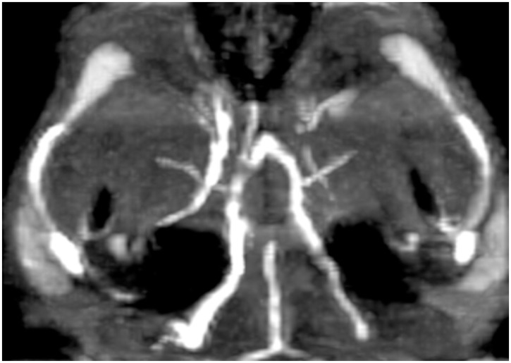
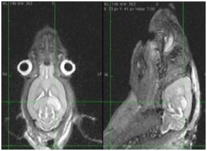
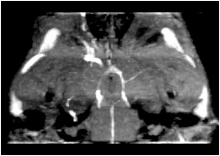
SAH was induced using the double-hemorrhage model in 12 rats. In two rats, saline solution was injected instead of blood (sham group). MR was performed on day 1, 2 and on day 5. T1-, T2-, T2*-weighted and time-of-flight MR sequences were applied, which were analyzed by two blinded neuroradiologists. Vessel narrowing of 25-50% was defined as mild, 50-75% as moderate and >75% as severe VSP.

RESULTS

We performed a total of 34 MRAs in 14 rats. In 14 rats, MRA was performed on day 2 and day 5. In six rats MRA was additionally performed on day1 before the blood injection. A good visualization of cerebral vessels was possible in all cases. No VSP was seen in the sham group neither on day 2 nor on day 5. We found vasospasm on day 2 in 7 of the 14 rats (50%) whereas all 7 rats had mild and one rat had additionally moderate and severe vasospasm in one vessel, respectively. On day 5 we found vasospasm in 8 of the 14 rats (60%) whereas 4 rats had severe vasospasm, 1 rat had moderate vasospasm and 3 rats demonstrated mild vasospasm. In 4 of the 14 rats (30%) an ischemic lesion was detected on day 5. Three of these rats had severe vasospasm and one rat had mild vasospasm. Severe vasospasm on day 5 was statistically significant correlated with the occurrence of ischemic lesions (Fisher's Exact test, OR 19.5, p = 0.03).

CONCLUSIONS

MRA is a noninvasive diagnostic tool, which allows a good visualization of the cerebral vasculature and provides reproducible results concerning the detection of VSP and the differentiation into three severity grades in rats. Future studies are needed to directly compare MRA with conventional angiography.

摘要

目的

磁共振(MR)成像已用于检测大鼠实验性蛛网膜下腔出血(eSAH)相关的脑血管痉挛(VSP)梗死。传统血管造影术通常用于观察VSP,这是一种侵入性技术,可能会增加发病率和死亡率。在本研究中,我们评估了磁共振血管造影(MRA)在检测大鼠eSAH后VSP的有效性及其定义VSP严重程度分级的可行性。

方法

采用双次出血模型诱导12只大鼠发生SAH。在2只大鼠中,注射生理盐水而非血液(假手术组)。在第1天、第2天和第5天进行MR检查。应用T1加权、T2加权、T2*加权和飞行时间MR序列,由两名不知情的神经放射科医生进行分析。血管狭窄25%-50%定义为轻度VSP,50%-75%为中度VSP,>75%为重度VSP。

结果

我们对14只大鼠共进行了34次MRA检查。14只大鼠在第2天和第5天进行了MRA检查。6只大鼠在注射血液前的第1天额外进行了MRA检查。所有病例均能很好地显示脑血管。假手术组在第2天和第5天均未发现VSP。我们发现14只大鼠中有7只(50%)在第2天出现血管痉挛,其中所有7只大鼠均为轻度血管痉挛,1只大鼠在一根血管中还出现了中度和重度血管痉挛。在第5天,我们发现14只大鼠中有8只(60%)出现血管痉挛,其中4只大鼠为重度血管痉挛,1只大鼠为中度血管痉挛,3只大鼠为轻度血管痉挛。14只大鼠中有4只(30%)在第5天检测到缺血性病变。其中3只大鼠有重度血管痉挛,1只大鼠有轻度血管痉挛。第5天的重度血管痉挛与缺血性病变的发生在统计学上有显著相关性(Fisher精确检验,OR 19.5,p = 0.03)。

结论

MRA是一种非侵入性诊断工具,能够很好地显示脑血管系统,并在检测大鼠VSP及将其分为三个严重程度等级方面提供可重复的结果。未来需要进行研究以直接比较MRA与传统血管造影术。

https://cdn.ncbi.nlm.nih.gov/pmc/blobs/7e57/5300130/b8ae7b8914d3/pone.0171121.g003.jpg
https://cdn.ncbi.nlm.nih.gov/pmc/blobs/7e57/5300130/9d609f4342db/pone.0171121.g001.jpg
https://cdn.ncbi.nlm.nih.gov/pmc/blobs/7e57/5300130/1d80a60a258e/pone.0171121.g002.jpg
https://cdn.ncbi.nlm.nih.gov/pmc/blobs/7e57/5300130/b8ae7b8914d3/pone.0171121.g003.jpg
https://cdn.ncbi.nlm.nih.gov/pmc/blobs/7e57/5300130/9d609f4342db/pone.0171121.g001.jpg
https://cdn.ncbi.nlm.nih.gov/pmc/blobs/7e57/5300130/1d80a60a258e/pone.0171121.g002.jpg
https://cdn.ncbi.nlm.nih.gov/pmc/blobs/7e57/5300130/b8ae7b8914d3/pone.0171121.g003.jpg

相似文献

1
MR-angiography allows defining severity grades of cerebral vasospasm in an experimental double blood injection subarachnoid hemorrhage model in rats.磁共振血管造影术能够在大鼠实验性双血注射蛛网膜下腔出血模型中确定脑血管痉挛的严重程度分级。
PLoS One. 2017 Feb 9;12(2):e0171121. doi: 10.1371/journal.pone.0171121. eCollection 2017.
2
Three-dimensional spin-echo-based black-blood MRA in the detection of vasospasm following subarachnoid hemorrhage.基于三维自旋回波的黑血磁共振血管成像在蛛网膜下腔出血后血管痉挛检测中的应用。
J Magn Reson Imaging. 2019 Mar;49(3):800-807. doi: 10.1002/jmri.26231. Epub 2018 Oct 4.
3
Canine double hemorrhage model of experimental subarachnoid hemorrhage.犬实验性蛛网膜下腔出血双出血模型
Acta Neurochir Suppl. 2015;120:347-51. doi: 10.1007/978-3-319-04981-6_60.
4
Time course in the development of cerebral vasospasm after experimental subarachnoid hemorrhage: clinical and neuroradiological assessment of the rat double hemorrhage model.实验性蛛网膜下腔出血后脑血管痉挛发展的时间进程:大鼠双重出血模型的临床和神经放射学评估
Neurosurgery. 2006 Jun;58(6):1190-7; discussion 1190-7. doi: 10.1227/01.NEU.0000199346.74649.66.
5
Quantitative magnetic resonance angiography as an alternative imaging technique in the assessment of cerebral vasospasm after subarachnoid hemorrhage.定量磁共振血管造影在蛛网膜下腔出血后脑血管痉挛评估中的替代成像技术。
Interv Neuroradiol. 2024 Apr;30(2):271-279. doi: 10.1177/15910199221138167. Epub 2022 Nov 10.
6
A low mortality rat model to assess delayed cerebral vasospasm after experimental subarachnoid hemorrhage.一种用于评估实验性蛛网膜下腔出血后迟发性脑血管痉挛的低死亡率大鼠模型。
J Vis Exp. 2013 Jan 17(71):e4157. doi: 10.3791/4157.
7
Non-invasive assessment of vasospasm following aneurysmal SAH using C-arm FDCT parenchymal blood volume measurement in the neuro-interventional suite: Technical feasibility.在神经介入手术室中使用C型臂FDCT实质血容量测量对动脉瘤性蛛网膜下腔出血后的血管痉挛进行无创评估:技术可行性
Interv Neuroradiol. 2015 Aug;21(4):479-89. doi: 10.1177/1591019915582376. Epub 2015 May 27.
8
Clinical utility of multislice computed tomographic angiography for detection of cerebral vasospasm in acute subarachnoid hemorrhage.多层螺旋计算机断层血管造影术在急性蛛网膜下腔出血中检测脑血管痉挛的临床应用价值
Minim Invasive Neurosurg. 2006 Oct;49(5):286-90. doi: 10.1055/s-2006-954826.
9
Double cisterna magna blood injection model of experimental subarachnoid hemorrhage in dogs.犬实验性蛛网膜下腔出血的双枕大池注血模型
Transl Stroke Res. 2014 Dec;5(6):647-52. doi: 10.1007/s12975-014-0356-8. Epub 2014 Jul 3.
10
Vasospasm after subarachnoid hemorrhage: diagnosis with MR angiography.蛛网膜下腔出血后的血管痉挛:磁共振血管造影诊断
AJNR Am J Neuroradiol. 2000 Oct;21(9):1611-7.

引用本文的文献

1
Advances in biomarkers for vasospasm - Towards a future blood-based diagnostic test.血管痉挛生物标志物的进展——迈向未来基于血液的诊断测试
World Neurosurg X. 2024 Mar 3;22:100343. doi: 10.1016/j.wnsx.2024.100343. eCollection 2024 Apr.
2
Delayed Cerebral Ischemia After Subarachnoid Hemorrhage: Is There a Relevant Experimental Model? A Systematic Review of Preclinical Literature.蛛网膜下腔出血后迟发性脑缺血:是否存在相关实验模型?临床前文献的系统评价
Front Cardiovasc Med. 2021 Nov 15;8:752769. doi: 10.3389/fcvm.2021.752769. eCollection 2021.
3
miR-24 targets HMOX1 to regulate inflammation and neurofunction in rats with cerebral vasospasm after subarachnoid hemorrhage.

本文引用的文献

1
Rat endovascular perforation model.大鼠血管内穿孔模型。
Transl Stroke Res. 2014 Dec;5(6):660-8. doi: 10.1007/s12975-014-0368-4. Epub 2014 Sep 13.
2
Delayed cerebral ischaemia after subarachnoid haemorrhage: looking beyond vasospasm.蛛网膜下腔出血后的迟发性脑缺血:超越血管痉挛的思考。
Br J Anaesth. 2012 Sep;109(3):315-29. doi: 10.1093/bja/aes264.
3
The effect of common carotid artery occlusion on delayed brain tissue damage in the rat double subarachnoid hemorrhage model.颈总动脉闭塞对大鼠双蛛网膜下腔出血模型迟发性脑组织损伤的影响。
微小RNA-24靶向血红素加氧酶-1以调节蛛网膜下腔出血后发生脑血管痉挛的大鼠的炎症反应和神经功能。
Am J Transl Res. 2021 Mar 15;13(3):1064-1074. eCollection 2021.
4
The impact of transcranial direct current stimulation on cerebral vasospasm in a rat model of subarachnoid hemorrhage.经颅直流电刺激对蛛网膜下腔出血大鼠模型脑血管痉挛的影响。
J Cereb Blood Flow Metab. 2021 Aug;41(8):2000-2009. doi: 10.1177/0271678X21990130. Epub 2021 Jan 27.
5
The ESAS-score: A histological severity grading system of subarachnoid hemorrhage using the modified double hemorrhage model in rats.ESAS 评分:一种使用改良大鼠二次出血模型的蛛网膜下腔出血组织学严重程度分级系统。
PLoS One. 2020 Feb 25;15(2):e0227349. doi: 10.1371/journal.pone.0227349. eCollection 2020.
Acta Neurochir (Wien). 2012 Jan;154(1):11-9. doi: 10.1007/s00701-011-1191-2. Epub 2011 Oct 11.
4
Effect of pharmaceutical treatment on vasospasm, delayed cerebral ischemia, and clinical outcome in patients with aneurysmal subarachnoid hemorrhage: a systematic review and meta-analysis.药物治疗对颅内动脉瘤性蛛网膜下腔出血患者血管痉挛、迟发性脑缺血和临床转归的影响:系统评价和荟萃分析。
J Cereb Blood Flow Metab. 2011 Jun;31(6):1443-51. doi: 10.1038/jcbfm.2011.7. Epub 2011 Feb 2.
5
A new approach to the treatment of cerebral vasospasm: the angiographic effects of tadalafil on experimental vasospasm.一种新的治疗脑血管痉挛的方法:他达拉非对实验性血管痉挛的血管造影效果。
Acta Neurochir (Wien). 2010 Mar;152(3):463-9. doi: 10.1007/s00701-009-0540-x. Epub 2009 Oct 20.
6
Comparison of experimental rat models of early brain injury after subarachnoid hemorrhage.蛛网膜下腔出血后早期脑损伤实验大鼠模型的比较
Neurosurgery. 2009 Aug;65(2):331-43; discussion 343. doi: 10.1227/01.NEU.0000345649.78556.26.
7
Clazosentan to overcome neurological ischemia and infarction occurring after subarachnoid hemorrhage (CONSCIOUS-1): randomized, double-blind, placebo-controlled phase 2 dose-finding trial.克拉生坦治疗蛛网膜下腔出血后发生的神经缺血和梗死(CONSCIOUS-1):随机、双盲、安慰剂对照2期剂量探索试验
Stroke. 2008 Nov;39(11):3015-21. doi: 10.1161/STROKEAHA.108.519942. Epub 2008 Aug 7.
8
Time course in the development of cerebral vasospasm after experimental subarachnoid hemorrhage: clinical and neuroradiological assessment of the rat double hemorrhage model.实验性蛛网膜下腔出血后脑血管痉挛发展的时间进程:大鼠双重出血模型的临床和神经放射学评估
Neurosurgery. 2006 Jun;58(6):1190-7; discussion 1190-7. doi: 10.1227/01.NEU.0000199346.74649.66.
9
Magnetic resonance imaging in experimental subarachnoid haemorrhage.实验性蛛网膜下腔出血的磁共振成像
Acta Neurochir (Wien). 2005 Sep;147(9):977-83; discussion 983. doi: 10.1007/s00701-005-0539-x. Epub 2005 May 25.
10
Complications and pitfalls in rat stroke models for middle cerebral artery occlusion: a comparison between the suture and the macrosphere model using magnetic resonance angiography.大鼠大脑中动脉闭塞性卒中模型的并发症与陷阱:使用磁共振血管造影术对缝合线模型和微球模型的比较
Stroke. 2004 Oct;35(10):2372-7. doi: 10.1161/01.STR.0000142134.37512.a7. Epub 2004 Sep 2.