Roberts Rosalinda C
Department of Psychiatry and Behavioral Neurobiology, University of Alabama, Birmingham, AL 35294, United States.
Schizophr Res. 2017 Sep;187:17-25. doi: 10.1016/j.schres.2017.01.056. Epub 2017 Feb 9.
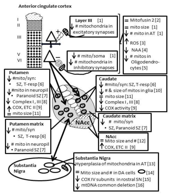
The aim of this paper is to provide a brief review of mitochondrial structure as it relates to function and then present abnormalities in mitochondria in postmortem schizophrenia with a focus on ultrastructure. Function, morphology, fusion, fission, motility, ΔΨmem, ATP production, mitochondrial derived vesicles, and mitochondria-associated ER membranes will be briefly covered. Pathology in mitochondria has long been implicated in schizophrenia, as shown by genetic, proteomic, enzymatic and anatomical abnormalities. The cortex and basal ganglia will be reviewed. In the anterior cingulate cortex, the number of mitochondria per neuronal somata in layers 5/6 in schizophrenia is decreased by 43%. There are also fewer mitochondria in terminals forming axospinous synapses. In the caudate and putamen the number of mitochondria is abnormal in both glial cells and neurons in schizophrenia subjects, the extent of which depends on treatment, response and predominant lifetime symptoms. Treatment-responsive schizophrenia subjects had about a 40% decrease in the number of mitochondria per synapse in the caudate nucleus and putamen, while treatment resistant cases had normal values. A decrease in mitochondrial density in the neuropil distinguishes paranoid from undifferentiated schizophrenia. The appearance, size and density of mitochondria were normal in the nucleus accumbens. In the substantia nigra, COX subunits were affected in rostral regions. Mitochondrial hyperplasia occurs within axon terminals that synapse onto dopamine neurons, but mitochondria in dopamine neuronal somata are similar in size and number. In schizophrenia, mitochondria are differentially affected depending on the brain region, cell type, subcellular location, treatment status, treatment response and symptoms.
本文旨在简要综述与功能相关的线粒体结构,然后重点阐述死后精神分裂症中线粒体的异常情况,特别是超微结构方面。将简要涵盖线粒体的功能、形态、融合、裂变、运动性、膜电位、ATP生成、线粒体衍生囊泡以及线粒体相关内质网膜。线粒体病理学长期以来一直被认为与精神分裂症有关,这已通过遗传、蛋白质组学、酶学和解剖学异常得到证实。本文将对大脑皮层和基底神经节进行综述。在扣带回前部皮层,精神分裂症患者5/6层神经元胞体中线粒体数量减少了43%。形成轴棘突触的终末中的线粒体也较少。在尾状核和壳核中,精神分裂症患者的神经胶质细胞和神经元中的线粒体数量均异常,其异常程度取决于治疗、反应和主要的终生症状。治疗反应良好的精神分裂症患者尾状核和壳核中每个突触的线粒体数量减少约40%,而治疗抵抗的病例则为正常数值。神经毡中线粒体密度降低可区分偏执型和未分化型精神分裂症。伏隔核中线粒体的外观、大小和密度均正常。在黑质中,喙部区域的细胞色素氧化酶亚基受到影响。在与多巴胺能神经元形成突触的轴突终末内发生线粒体增生,但多巴胺能神经元胞体中的线粒体大小和数量相似。在精神分裂症中,线粒体根据脑区、细胞类型、亚细胞位置、治疗状态、治疗反应和症状的不同而受到不同影响。