Valarmathi Mani T, Fuseler John W, Davis Jeffrey M, Price Robert L
Laboratory of Stem Cell Biology and Tissue Engineering, Department of Comparative Biosciences, College of Veterinary Medicine, University of Illinois at Urbana-Champaign Urbana, IL, USA.
Department of Pathology, Microbiology and Immunology, School of Medicine, University of South Carolina Columbia, SC, USA.
Front Cell Dev Biol. 2017 Jan 30;5:2. doi: 10.3389/fcell.2017.00002. eCollection 2017.
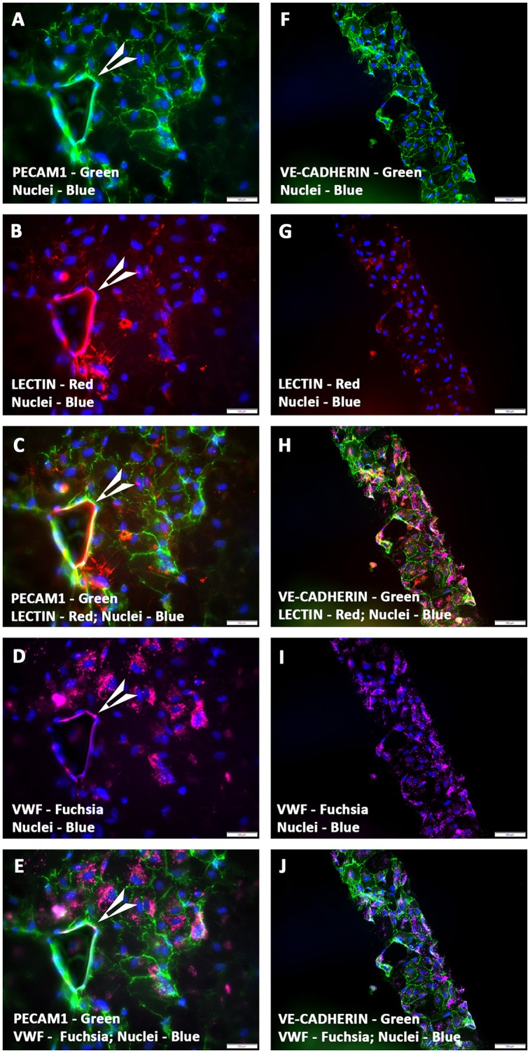
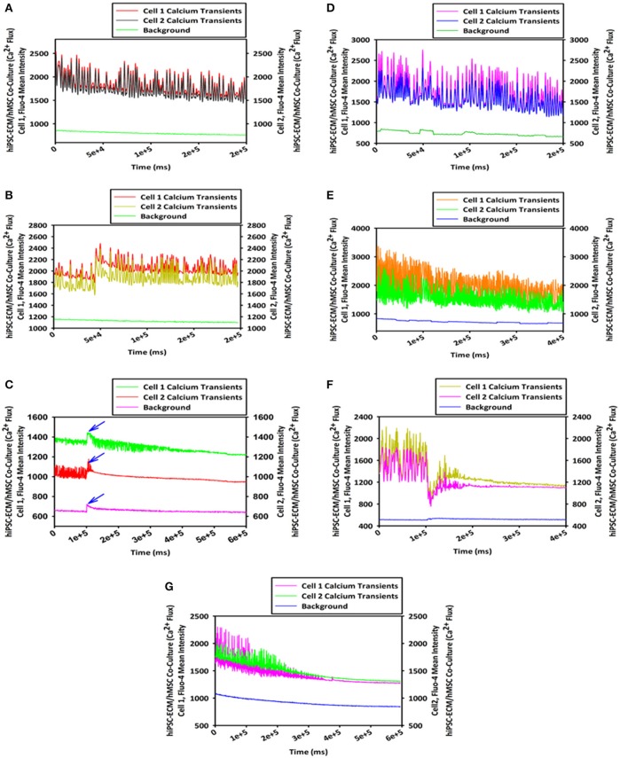
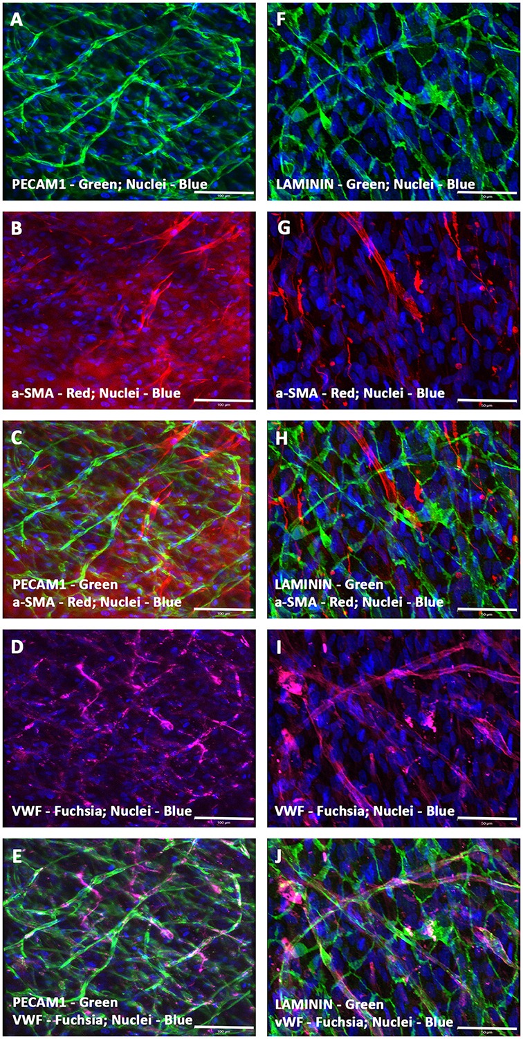
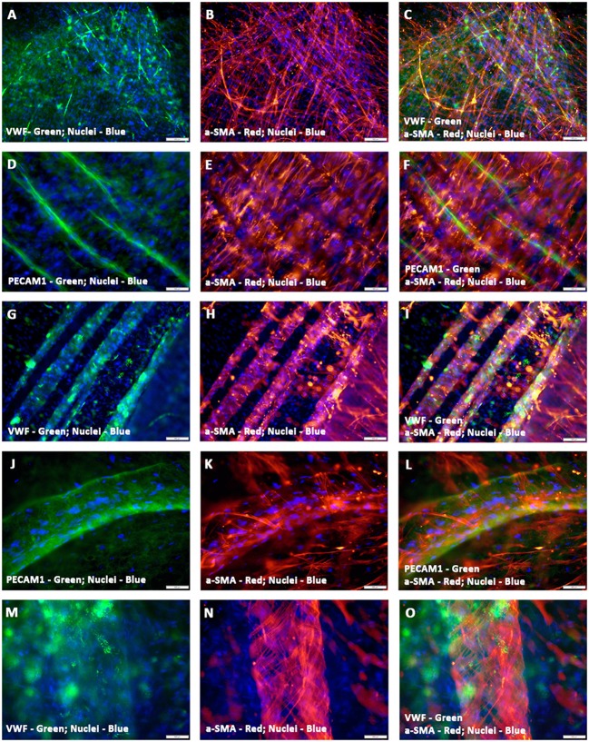
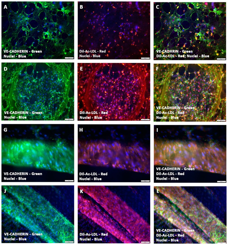
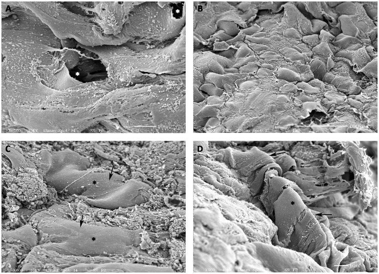
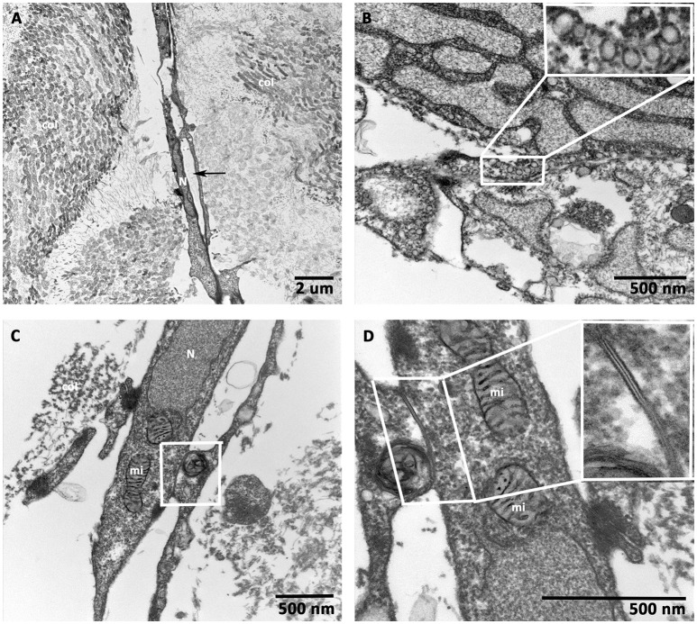
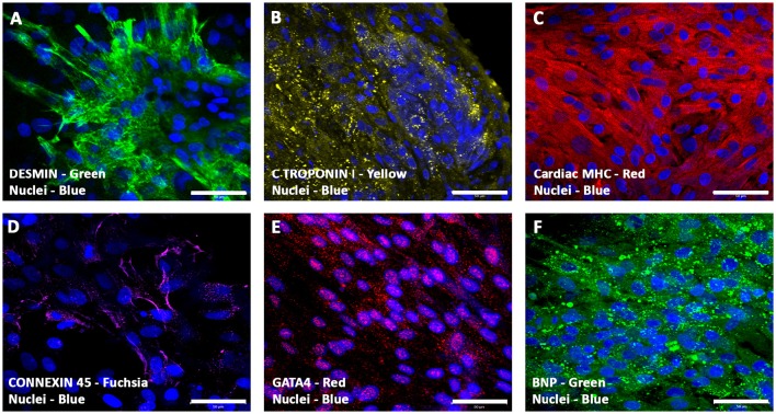
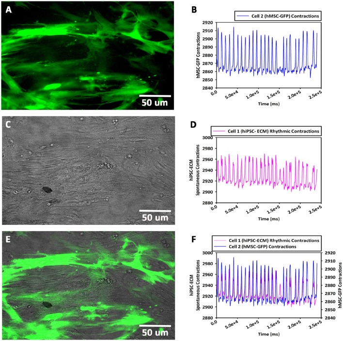
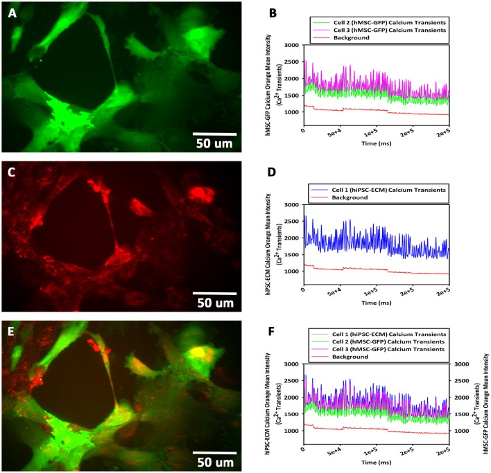
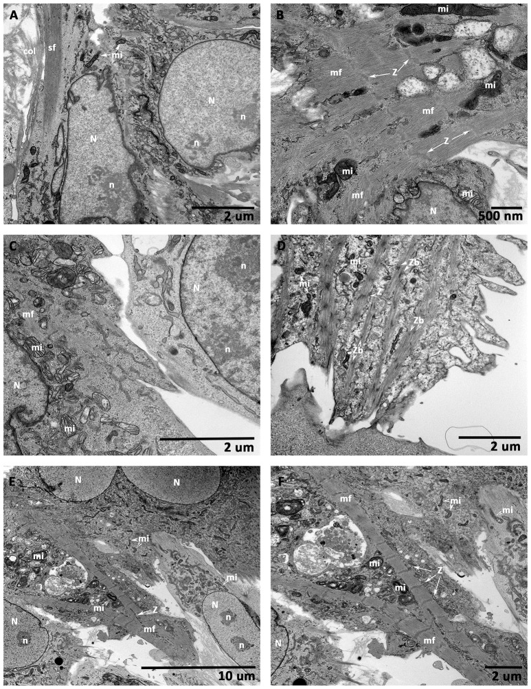
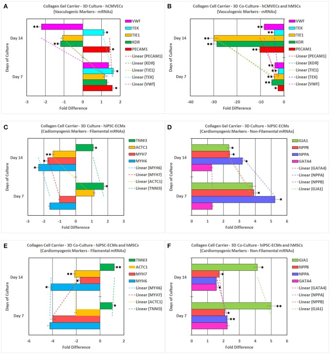
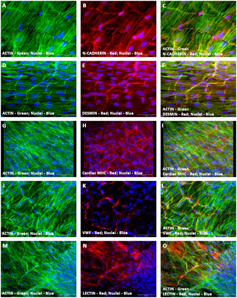
Organ tissue engineering, including cardiovascular tissues, has been an area of intense investigation. The major challenge to these approaches has been the inability to vascularize and perfuse the engineered tissue constructs. Attempts to provide oxygen and nutrients to the cells contained in the biomaterial constructs have had varying degrees of success. The aim of this current study is to develop a three-dimensional (3-D) model of vascularized cardiac tissue to examine the concurrent temporal and spatial regulation of cardiomyogenesis in the context of postnatal vasculogenesis during stem cell cardiac regeneration. In order to achieve the above aim, we have developed an 3-D functional vascularized cardiac muscle construct using human induced pluripotent stem cell-derived embryonic cardiac myocytes (hiPSC-ECMs) and human mesenchymal stem cells (hMSCs). First, to generate the prevascularized scaffold, human cardiac microvascular endothelial cells (hCMVECs) and hMSCs were co-cultured onto a 3-D collagen cell carrier (CCC) for 7 days under vasculogenic culture conditions. In this milieu, hCMVECs/hMSCs underwent maturation, differentiation, and morphogenesis characteristic of microvessels, and formed extensive plexuses of vascular networks. Next, the hiPSC-ECMs and hMSCs were co-cultured onto this generated prevascularized CCCs for further 7 or 14 days in myogenic culture conditions. Finally, the vascular and cardiac phenotypic inductions were analyzed at the morphological, immunological, biochemical, molecular, and functional levels. Expression and functional analyses of the differentiated cells revealed neo-angiogenesis and neo-cardiomyogenesis. Thus, our unique 3-D co-culture system provided us the apt functional vascularized 3-D cardiac patch that can be utilized for cellular cardiomyoplasty.
包括心血管组织在内的器官组织工程一直是一个深入研究的领域。这些方法面临的主要挑战是无法使工程化组织构建体血管化并进行灌注。为生物材料构建体中所含细胞提供氧气和营养物质的尝试取得了不同程度的成功。本研究的目的是建立一种血管化心脏组织的三维(3-D)模型,以研究干细胞心脏再生过程中出生后血管生成背景下心肌发生的同时时空调节。为了实现上述目标,我们使用人诱导多能干细胞衍生的胚胎心肌细胞(hiPSC-ECM)和人间充质干细胞(hMSC)开发了一种三维功能性血管化心肌构建体。首先,为了生成预血管化支架,将人心脏微血管内皮细胞(hCMVEC)和hMSC在血管生成培养条件下共培养在三维胶原细胞载体(CCC)上7天。在这种环境中,hCMVEC/hMSC经历了微血管特有的成熟、分化和形态发生,并形成了广泛的血管网络丛。接下来,将hiPSC-ECM和hMSC在肌源性培养条件下共培养在这种生成的预血管化CCC上7天或14天。最后,在形态学、免疫学、生物化学、分子和功能水平上分析血管和心脏表型诱导。分化细胞的表达和功能分析显示了新血管生成和新心肌生成。因此,我们独特的三维共培养系统为我们提供了适合用于细胞心肌成形术的功能性血管化三维心脏补片。