Iraha Rin, Tsuchiya Nanae, Yamashiro Tsuneo, Iwasawa Tae, Murayama Sadayuki
Department of Radiology, Graduate School of Medical Science, University of the Ryukyus, Okinawa, Japan.
Department of Radiology, Kanagawa Cardiovascular and Respiratory Center, Kanagawa, Japan.
Acta Radiol Open. 2017 Jan 1;6(1):2058460116684370. doi: 10.1177/2058460116684370. eCollection 2017 Jan.
Magnetic resonance imaging (MRI) can be beneficial for diagnosis of disease by offering quantitative information. However, reproducibility can be a major problem when there is a numerical threshold in multi-institution, multi-vendor situations.
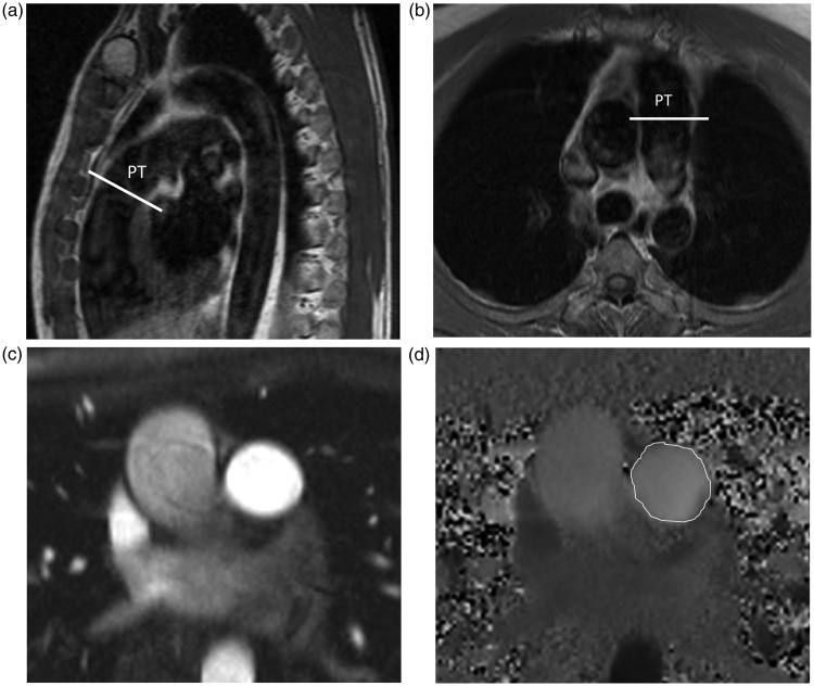
To measure pulmonary blood flow with phase-contrast (PC) imaging using two different MR scanners (1.5 T) at different institutions in the same participants and to examine the reproducibility of the measurements.
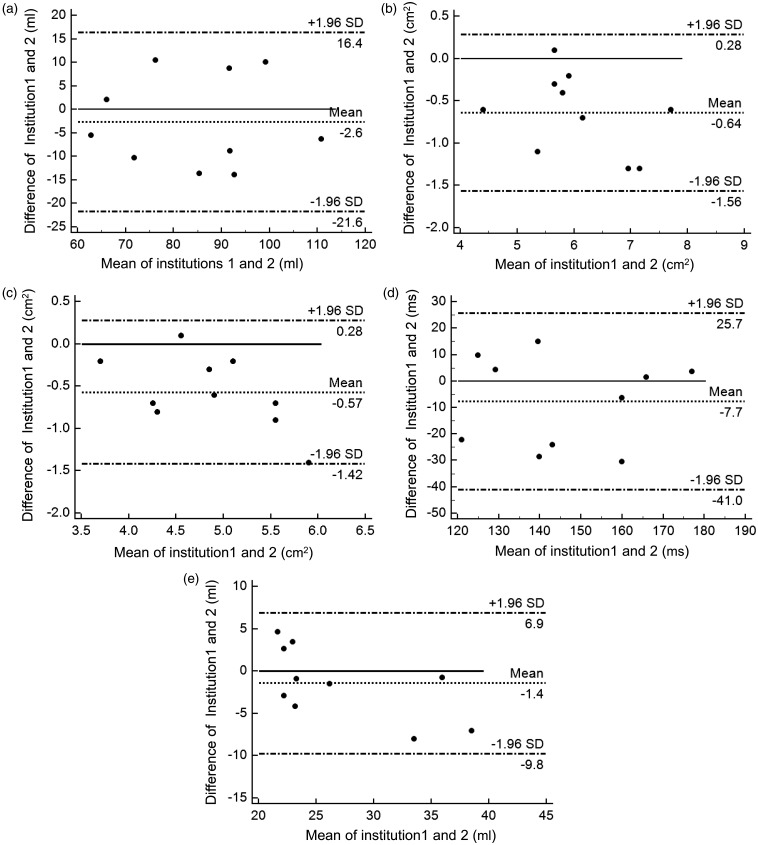
Participants were 10 healthy volunteers (5 men; age range, 27-36 years). The measurements included the mean and maximal blood velocities, the mean blood flow volume, and the acceleration time and volume (AT and AV), derived from the time-flow curve of the PC-MRI. Simultaneously obtained maximal, minimal, and mean areas from regions of interest set in the pulmonary artery were also calculated. In order to calculate the reproducibility of the quantitative variables, intra-class correlation coefficients (ICCs) were employed. When an adequate ICC was obtained, Bland-Altman analysis was conducted to identify any systematic bias.
The ICCs were almost perfect for the mean blood flow volume and the AV (r = 0.82 and 0.80), and were substantial in the mean and maximal areas, and the AT (r = 0.63, 0.74, and 0.64, respectively). However, there was a fixed bias in the area measurement between the two scanners. Also, the AV had a proportional bias.
Our results reveal that various indices derived from PC-MRI on different MR scanners are promising as common indices for pulmonary flow assessment. Research and clinical use of PC-MRI for the pulmonary artery is expected to extend to multi-institution situations.
磁共振成像(MRI)通过提供定量信息有助于疾病诊断。然而,在多机构、多供应商的情况下,当存在数值阈值时,可重复性可能是一个主要问题。
在同一参与者的不同机构使用两台不同的磁共振扫描仪(1.5T),通过相位对比(PC)成像测量肺血流量,并检验测量结果的可重复性。
参与者为10名健康志愿者(5名男性;年龄范围27 - 36岁)。测量内容包括平均和最大血流速度、平均血流量、加速时间和加速容积(AT和AV),这些数据来自PC - MRI的时间 - 血流曲线。同时还计算了在肺动脉中设置的感兴趣区域的最大、最小和平均面积。为了计算定量变量的可重复性,采用组内相关系数(ICC)。当获得足够的ICC时,进行布兰德 - 奥特曼分析以识别任何系统偏差。
平均血流量和AV的ICC几乎为完美(r = 0.82和0.80),平均和最大面积以及AT的ICC为实质性相关(分别为r = 0.63、0.74和0.64)。然而,两台扫描仪之间的面积测量存在固定偏差。此外,AV存在比例偏差。
我们的结果表明,不同磁共振扫描仪上PC - MRI得出的各种指标有望成为肺血流评估的通用指标。肺动脉PC - MRI的研究和临床应用有望扩展到多机构情况。