Dieterich Lothar C, Ikenberg Kristian, Cetintas Timur, Kapaklikaya Kübra, Hutmacher Cornelia, Detmar Michael
Institute of Pharmaceutical Sciences, Swiss Federal Institute of Technology (ETH) Zurich , Zurich , Switzerland.
Institute of Pharmaceutical Sciences, Swiss Federal Institute of Technology (ETH) Zurich, Zurich, Switzerland; Department of Pathology and Molecular Pathology, University Hospital Zurich, Zurich, Switzerland.
Front Immunol. 2017 Feb 3;8:66. doi: 10.3389/fimmu.2017.00066. eCollection 2017.
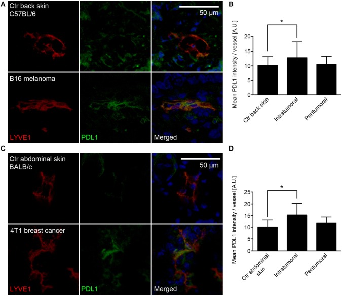
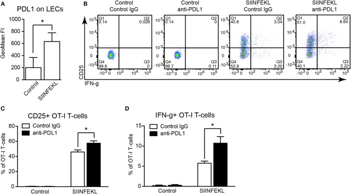
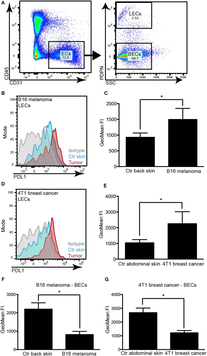
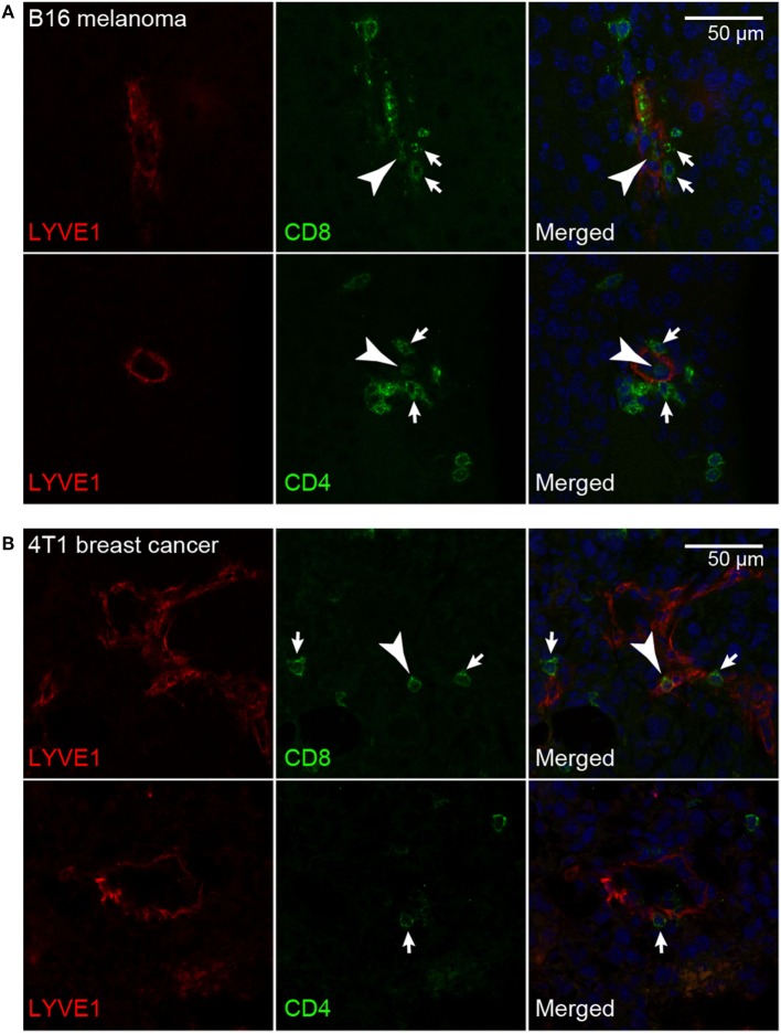
Tumor-associated lymphatic vessels (LVs) play multiple roles during tumor progression, including promotion of metastasis and regulation of antitumor immune responses by delivering antigen from the tumor bed to draining lymph nodes (LNs). Under steady-state conditions, LN resident lymphatic endothelial cells (LECs) have been found to maintain peripheral tolerance by directly inhibiting autoreactive T-cells. Similarly, tumor-associated lymphatic endothelium has been suggested to reduce antitumor T-cell responses, but the mechanisms that mediate this effect have not been clarified. Using two distinct experimental tumor models, we found that tumor-associated LVs gain expression of the T-cell inhibitory molecule PDL1, similar to LN resident LECs, whereas tumor-associated blood vessels downregulate PDL1. The observed lymphatic upregulation of PDL1 was likely due to IFN-g released by stromal cells in the tumor microenvironment. Furthermore, we found that blocking PDL1 results in increased T-cell stimulation by antigen-presenting LECs . Taken together, our data suggest that peripheral, tumor-associated lymphatic endothelium contributes to T-cell inhibition, by a mechanism similar to peripheral tolerance maintenance described for LN resident LECs. These findings may have clinical implications for cancer therapy, as lymphatic expression of PDL1 could represent a new biomarker to select patients for immunotherapy with PD1 or PDL1 inhibitors.
肿瘤相关淋巴管(LVs)在肿瘤进展过程中发挥多种作用,包括通过将抗原从肿瘤床输送至引流淋巴结(LNs)来促进转移和调节抗肿瘤免疫反应。在稳态条件下,已发现淋巴结驻留淋巴管内皮细胞(LECs)通过直接抑制自身反应性T细胞来维持外周耐受。同样,有人提出肿瘤相关淋巴管内皮可降低抗肿瘤T细胞反应,但介导这种效应的机制尚未阐明。使用两种不同的实验性肿瘤模型,我们发现肿瘤相关LVs获得了T细胞抑制分子PDL1的表达,类似于淋巴结驻留LECs,而肿瘤相关血管则下调PDL1。观察到的淋巴管中PDL1的上调可能是由于肿瘤微环境中基质细胞释放的IFN-γ所致。此外,我们发现阻断PDL1会导致抗原呈递LECs对T细胞的刺激增加。综上所述,我们的数据表明,外周肿瘤相关淋巴管内皮通过一种类似于淋巴结驻留LECs维持外周耐受的机制,促进T细胞抑制。这些发现可能对癌症治疗具有临床意义,因为PDL1的淋巴管表达可能代表一种新的生物标志物,用于选择接受PD1或PDL1抑制剂免疫治疗的患者。