Castro B A, Flanigan P, Jahangiri A, Hoffman D, Chen W, Kuang R, De Lay M, Yagnik G, Wagner J R, Mascharak S, Sidorov M, Shrivastav S, Kohanbash G, Okada H, Aghi M K
Department of Neurological Surgery, University of California San Francisco (UCSF), San Francisco, USA.
Oncogene. 2017 Jun 29;36(26):3749-3759. doi: 10.1038/onc.2017.1. Epub 2017 Feb 20.
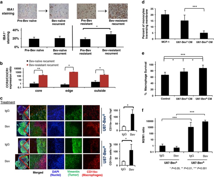
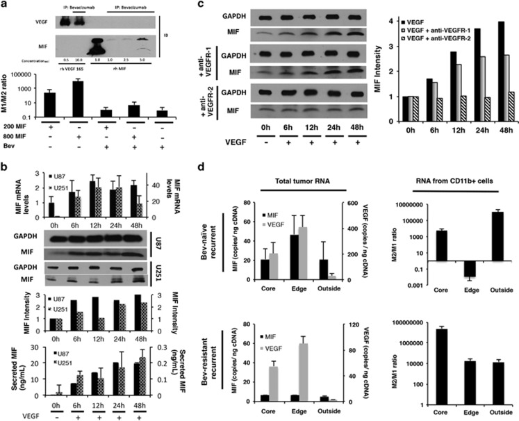
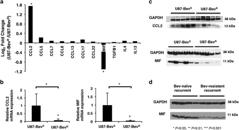
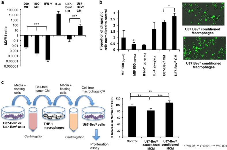
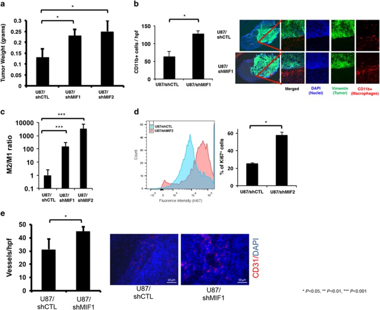
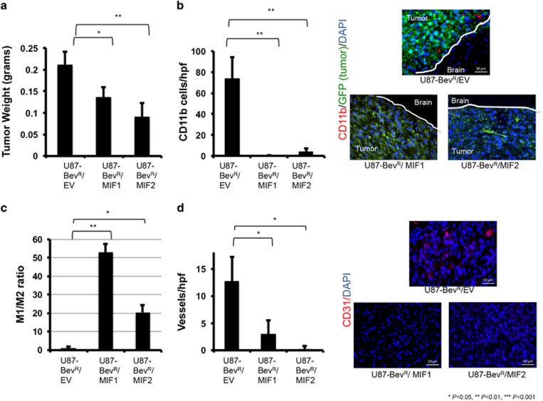
Anti-angiogenic therapies for cancer such as VEGF neutralizing antibody bevacizumab have limited durability. While mechanisms of resistance remain undefined, it is likely that acquired resistance to anti-angiogenic therapy will involve alterations of the tumor microenvironment. We confirmed increased tumor-associated macrophages in bevacizumab-resistant glioblastoma patient specimens and two novel glioblastoma xenograft models of bevacizumab resistance. Microarray analysis suggested downregulated macrophage migration inhibitory factor (MIF) to be the most pertinent mediator of increased macrophages. Bevacizumab-resistant patient glioblastomas and both novel xenograft models of resistance had less MIF than bevacizumab-naive tumors, and harbored more M2/protumoral macrophages that specifically localized to the tumor edge. Xenografts expressing MIF-shRNA grew more rapidly with greater angiogenesis and had macrophages localizing to the tumor edge which were more prevalent and proliferative, and displayed M2 polarization, whereas bevacizumab-resistant xenografts transduced to upregulate MIF exhibited the opposite changes. Bone marrow-derived macrophage were polarized to an M2 phenotype in the presence of condition-media derived from bevacizumab-resistant xenograft-derived cells, while recombinant MIF drove M1 polarization. Media from macrophages exposed to bevacizumab-resistant tumor cell conditioned media increased glioma cell proliferation compared with media from macrophages exposed to bevacizumab-responsive tumor cell media, suggesting that macrophage polarization in bevacizumab-resistant xenografts is the source of their aggressive biology and results from a secreted factor. Two mechanisms of bevacizumab-induced MIF reduction were identified: (1) bevacizumab bound MIF and blocked MIF-induced M1 polarization of macrophages; and (2) VEGF increased glioma MIF production in a VEGFR2-dependent manner, suggesting that bevacizumab-induced VEGF depletion would downregulate MIF. Site-directed biopsies revealed enriched MIF and VEGF at the enhancing edge in bevacizumab-naive patients. This MIF enrichment was lost in bevacizumab-resistant glioblastomas, driving a tumor edge M1-to-M2 transition. Thus, bevacizumab resistance is driven by reduced MIF at the tumor edge causing proliferative expansion of M2 macrophages, which in turn promotes tumor growth.
用于癌症的抗血管生成疗法,如VEGF中和抗体贝伐单抗,其疗效持久性有限。虽然耐药机制尚不清楚,但抗血管生成疗法的获得性耐药可能涉及肿瘤微环境的改变。我们在贝伐单抗耐药的胶质母细胞瘤患者标本以及两种新的贝伐单抗耐药胶质母细胞瘤异种移植模型中证实了肿瘤相关巨噬细胞增多。微阵列分析表明,巨噬细胞迁移抑制因子(MIF)下调是巨噬细胞增多的最相关介质。贝伐单抗耐药的患者胶质母细胞瘤以及两种新的耐药异种移植模型中的MIF均少于未接受过贝伐单抗治疗的肿瘤,并且含有更多特异性定位于肿瘤边缘的M2/促肿瘤巨噬细胞。表达MIF-shRNA的异种移植瘤生长更快,血管生成增加,巨噬细胞定位于肿瘤边缘,这些巨噬细胞更普遍且增殖,表现出M2极化,而转导上调MIF的贝伐单抗耐药异种移植瘤则表现出相反的变化。在存在来自贝伐单抗耐药异种移植瘤衍生细胞的条件培养基的情况下,骨髓来源的巨噬细胞极化为M2表型,而重组MIF驱动M1极化。与暴露于贝伐单抗敏感肿瘤细胞培养基的巨噬细胞的培养基相比,暴露于贝伐单抗耐药肿瘤细胞条件培养基的巨噬细胞的培养基增加了胶质瘤细胞的增殖,这表明贝伐单抗耐药异种移植瘤中的巨噬细胞极化是其侵袭性生物学行为的来源,并且是由一种分泌因子导致的。确定了贝伐单抗诱导MIF减少的两种机制:(1)贝伐单抗结合MIF并阻断MIF诱导的巨噬细胞M1极化;(2)VEGF以VEGFR2依赖的方式增加胶质瘤MIF的产生,这表明贝伐单抗诱导的VEGF耗竭会下调MIF。定点活检显示,在未接受过贝伐单抗治疗的患者中,增强边缘处的MIF和VEGF富集。在贝伐单抗耐药的胶质母细胞瘤中,这种MIF富集消失,导致肿瘤边缘从M1向M2转变。因此,贝伐单抗耐药是由肿瘤边缘MIF减少驱动的,MIF减少导致M2巨噬细胞增殖性扩张,进而促进肿瘤生长。