Suriyonplengsaeng Chinnawut, Dejthevaporn Charungthai, Khongkhatithum Chaiyos, Sanpapant Suda, Tubthong Nattha, Pinpradap Koset, Srinark Nippa, Waisayarat Jariya
Department of Pathology, Faculty of Medicine Ramathibodi Hospital, Mahidol University, Bangkok, 10400, Thailand.
Department of Anatomy, Faculty of Science, Mahidol University, Bangkok, 10400, Thailand.
Diagn Pathol. 2017 Feb 20;12(1):19. doi: 10.1186/s13000-017-0610-y.
The analysis of fresh frozen muscle specimens is standard following routine muscle biopsy, but this service is not widely available in countries with limited medical facilities, such as Thailand. Nevertheless, immunohistochemistry (IHC) analysis is essential for the diagnosis of patients with a strong clinical suspicion of muscular dystrophy, in the absence of mutations detected by molecular genetics. As the successful labelling of sarcolemmal membrane-associated proteins in formalin-fixed and paraffin-embedded (FFPE) muscle sections using IHC staining has rarely been described, this study aimed to develop a reproducible IHC method for such an analysis.
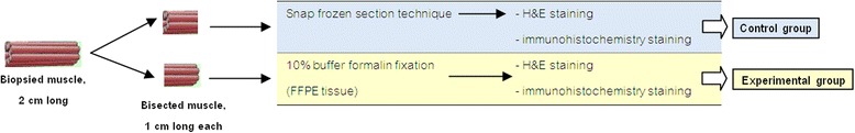
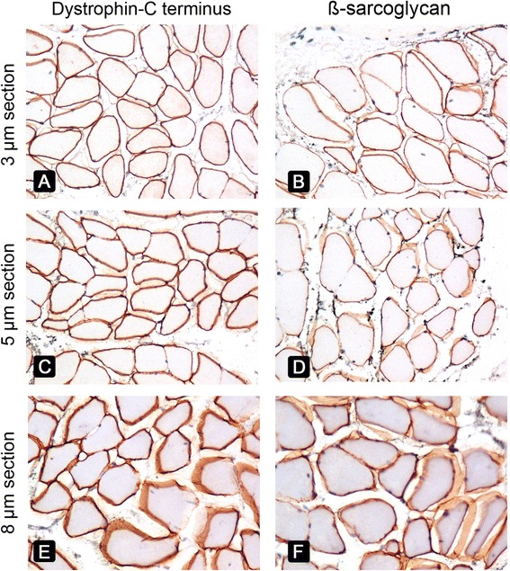
Thirteen cases were studied from the files of the Department of Pathology, Mahidol University. Diagnoses included three Duchenne muscular dystrophy (DMD), one Becker muscular dystrophy (BMD), one dysferlinopathy, and several not-specified muscular dystrophies. IHC was performed on FFPE sections at different thicknesses (3 μm, 5 μm, and 8 μm) using the heat-mediated antigen retrieval method with citrate/EDTA buffer, followed by an overnight incubation with primary antibodies at room temperature. Antibodies against spectrin, dystrophin (rod domain, C-terminus, and N-terminus), dysferlin, sarcoglycans (α, β, and γ), and β-dystroglycan were used. Frozen sections were tested in parallel for comparative analysis.
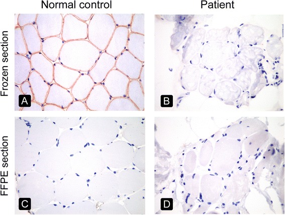
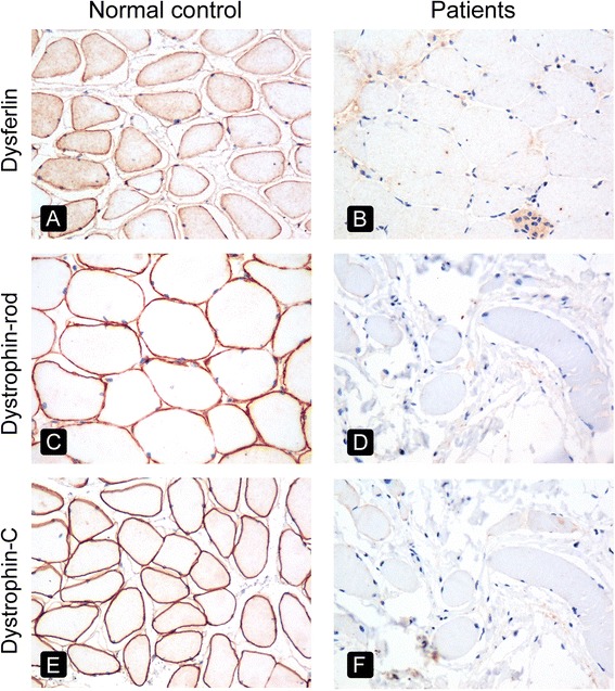
Antibodies labelling spectrin, dystrophin (rod domain and C-terminus), dysferlin, sarcoglycans (α, β, and γ), and β-dystroglycan clearly exhibited sarcolemmal staining in FFPE sections. However, staining of FFPE sections using the antibody directed against the N-terminus of dystrophin was unsuccessful. The absence of labeling for dystrophins and dysferlin in FFPE sections was documented in all three DMD patients and the dysferlinopathy patient. The BMD diagnosis could not be made using IHC in FFPE sections alone because of a lack of staining for the dystrophin N-terminus, indicating a limitation of this method.
We developed a reliable and reproducible IHC technique using FFPE muscle. This could become a valuable tool for the diagnosis of some muscular dystrophies, dystrophinopathies, sarcoglycanopathies (LGMD2D, LGMD2E, and LGMD2C), and dysferlinopathy, especially in situations where the analysis of fresh frozen muscle samples is not routinely available.
新鲜冷冻肌肉标本分析是常规肌肉活检后的标准操作,但在泰国等医疗设施有限的国家,这项服务并不广泛可用。然而,在分子遗传学未检测到突变的情况下,免疫组织化学(IHC)分析对于临床高度怀疑患有肌肉营养不良症的患者的诊断至关重要。由于很少有文献描述使用免疫组织化学染色在福尔马林固定石蜡包埋(FFPE)肌肉切片中成功标记肌膜相关蛋白,本研究旨在开发一种可重复的免疫组织化学方法用于此类分析。
从玛希隆大学病理学系的档案中选取13例病例进行研究。诊断包括3例杜氏肌营养不良症(DMD)、1例贝克肌营养不良症(BMD)、1例肌联蛋白病以及几例未明确诊断的肌肉营养不良症。使用柠檬酸盐/乙二胺四乙酸(EDTA)缓冲液的热介导抗原修复方法,对不同厚度(3μm、5μm和8μm) 的FFPE切片进行免疫组织化学检测,然后在室温下用一抗孵育过夜。使用了抗血影蛋白、抗肌营养不良蛋白(杆状结构域、C末端和N末端)、抗肌联蛋白、抗肌聚糖(α、β和γ)以及抗β - 肌营养不良聚糖的抗体。同时对冷冻切片进行检测以进行对比分析。
标记血影蛋白、肌营养不良蛋白(杆状结构域和C末端)、肌联蛋白、肌聚糖(α、β和γ)以及β - 肌营养不良聚糖的抗体在FFPE切片中清晰地显示出肌膜染色。然而,使用针对肌营养不良蛋白N末端的抗体对FFPE切片进行染色未成功。在所有3例DMD患者和肌联蛋白病患者的FFPE切片中,均记录到肌营养不良蛋白和肌联蛋白标记缺失。仅使用FFPE切片中的免疫组织化学方法无法做出BMD诊断,因为肌营养不良蛋白N末端缺乏染色,这表明了该方法的局限性。
我们开发了一种使用FFPE肌肉的可靠且可重复的免疫组织化学技术。这可能成为诊断某些肌肉营养不良症、肌营养不良蛋白病、肌聚糖病(LGMD2D、LGMD2E和LGMD2C)以及肌联蛋白病的有价值工具,特别是在无法常规获取新鲜冷冻肌肉样本进行分析的情况下。