Zhang Wei, Liu Xingzhou, Zuo Lijun, Guo Qiang, Chen Qi, Wang Yongjun
Department of Neurology, Beijing Tiantan Hospital, Capital Medical University, Beijing, China.
Epilepsy Center, Guangdong Sanjiu Brain Hospital, Jinan University, No. 578, Sha Tai Nan Lu, Guangzhou, 510510, China.
BMC Neurol. 2017 Feb 21;17(1):38. doi: 10.1186/s12883-017-0811-8.
Versive seizure characterized by conjugate eye movement during epileptic seizure has been considered commonly as one of the most valuable semiological signs for epilepsy localization, especially for frontal lobe epilepsy. However, the lateralizing and localizing significance of ictaleye deviation has been questioned by clinical observation of a series of focal epilepsy studies, including frontal, central, temporal, parietal and occipital epilepsy.
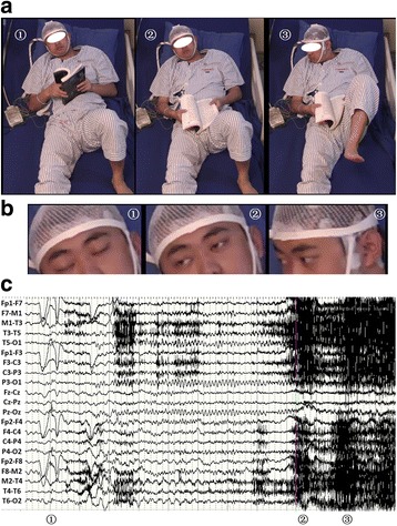
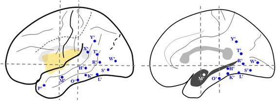
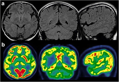
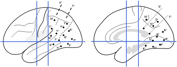
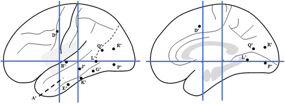
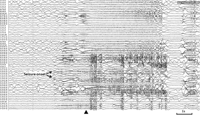
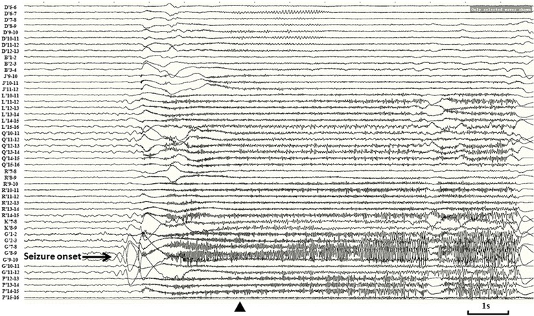
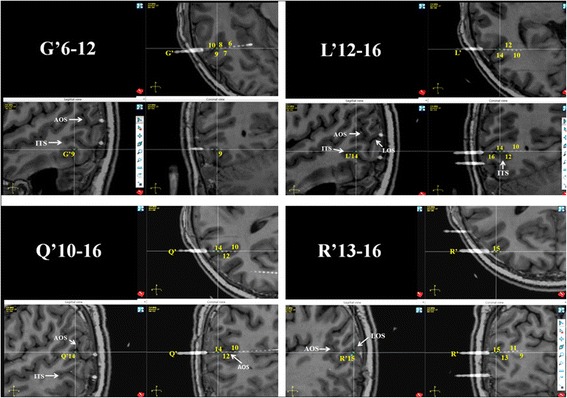
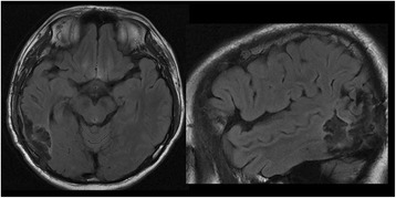
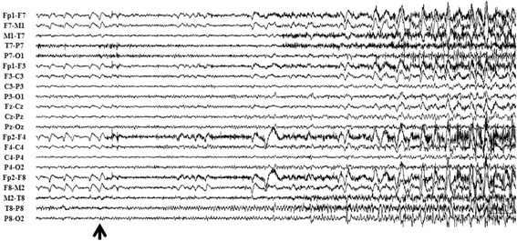
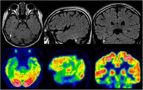
Two epileptic cases characterized by ipsiversive eye deviation as initial clinical sign during the habitual epileptic seizures are presented in this paper. The localization of the epileptogenic zone of both of the cases has been confirmed as inferioposterior temporal region by the findings of ictalstereoelectroencephalography (SEEG) and a good result after epileptic surgery. Detailed analysis of the exact position of the key contacts of the SEEG electrodes identified the overlap between the location of the epileptogenic zone and human MT/MST complex, which play a crucial role in the control of smooth pursuit eye movement.
Ipsiversive eye deviation could be the initial clinical sign of inferioposterior temporal lobe epilepsy and attribute to the involvement of human MT/MST complex, especially human MST whichwas located on the anterior/dorsal bank of the anterior occipital sulcus (AOS).
癫痫发作时以共轭眼球运动为特征的旋转性发作通常被认为是癫痫定位最有价值的半侧症状之一,尤其是对于额叶癫痫。然而,一系列局灶性癫痫研究(包括额叶、中央、颞叶、顶叶和枕叶癫痫)的临床观察对发作期眼球偏斜的定位和定侧意义提出了质疑。
本文介绍了两例癫痫病例,其习惯性癫痫发作期间以同侧旋转性眼球偏斜为初始临床症状。通过发作期立体脑电图(SEEG)检查结果证实两例患者的致痫区均位于颞叶后下部,癫痫手术后效果良好。对SEEG电极关键触点的确切位置进行详细分析,确定了致痫区位置与人类MT/MST复合体之间存在重叠,该复合体在控制平稳跟踪眼球运动中起关键作用。
同侧旋转性眼球偏斜可能是颞叶后下部癫痫的初始临床症状,这归因于人类MT/MST复合体的受累,尤其是位于枕前沟(AOS)前/背侧缘的人类MST。