• 文献检索
  • 文档翻译
  • 深度研究
  • 学术资讯
  • Suppr Zotero 插件Zotero 插件
  • 邀请有礼
  • 套餐&价格
  • 历史记录
应用&插件
Suppr Zotero 插件Zotero 插件浏览器插件Mac 客户端Windows 客户端微信小程序
定价
高级版会员购买积分包购买API积分包
服务
文献检索文档翻译深度研究API 文档MCP 服务
关于我们
关于 Suppr公司介绍联系我们用户协议隐私条款
关注我们

Suppr 超能文献

核心技术专利:CN118964589B侵权必究
粤ICP备2023148730 号-1Suppr @ 2026

文献检索

告别复杂PubMed语法,用中文像聊天一样搜索,搜遍4000万医学文献。AI智能推荐,让科研检索更轻松。

立即免费搜索

文件翻译

保留排版,准确专业,支持PDF/Word/PPT等文件格式,支持 12+语言互译。

免费翻译文档

深度研究

AI帮你快速写综述,25分钟生成高质量综述,智能提取关键信息,辅助科研写作。

立即免费体验

皮肤镜检查在破裂和未破裂表皮囊肿鉴别诊断中的应用价值

Usefulness of Dermoscopy in the Differential Diagnosis of Ruptured and Unruptured Epidermal Cysts.

作者信息

Suh Kee Suck, Kang Dong Young, Park Jong Bin, Yang Myeong Hyeon, Kim Joon Hee, Lee Kang Hoon, Han Sang Hwa, Choi Yun Deok, Kim Sang Tae, Jang Min Soo

机构信息

Department of Dermatology, Kosin University College of Medicine, Busan, Korea.

Miul Dermatologic Clinic, Busan, Korea.

出版信息

Ann Dermatol. 2017 Feb;29(1):33-38. doi: 10.5021/ad.2017.29.1.33. Epub 2017 Feb 3.

DOI:10.5021/ad.2017.29.1.33
PMID:28223744
原文链接:https://pmc.ncbi.nlm.nih.gov/articles/PMC5318524/
Abstract

BACKGROUND

An epidermal cyst is a common keratin-filled epithelial-lined cyst. The treatment of choice for epidermal cysts is surgical excision. If the cyst becomes ruptured, incision and drainage with oral antibiotic therapy or intralesional steroid injection are required.

OBJECTIVE

To analyze the dermoscopic features that can differentiate between ruptured and unruptured epidermal cysts.

METHODS

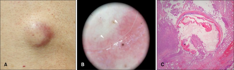
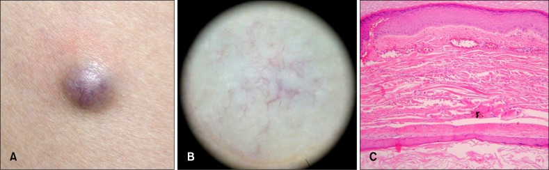
The clinical and dermoscopic features of the pathologically confirmed epidermal cysts of two subgroups of 38 patients, 20 with unruptured cysts and 18 with ruptured cysts, were reviewed.

RESULTS

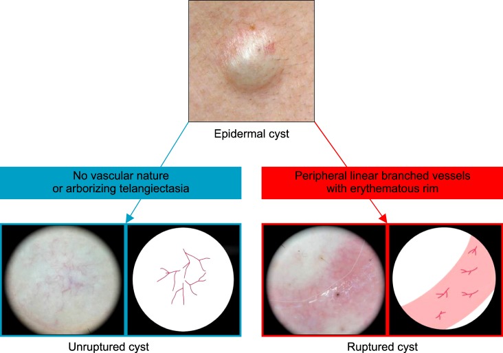
With regard to the dermoscopic features, an ivory- white background color and punctum were commonly found in both groups (>0.05). The unruptured-cyst group showed higher frequencies of pore sign (<0.05), blue-white veil (>0.05), no vascular structure, and arborizing telangiectasia (<0.05), but the ruptured-cyst group usually had red lacunae (>0.05) and peripheral linear branched vessels (with an erythematous rim) (<0.05).

CONCLUSION

Dermoscopy is helpful in differentiating between ruptured and unruptured epidermal cysts.

摘要

背景

表皮囊肿是一种常见的充满角蛋白的上皮内衬囊肿。表皮囊肿的首选治疗方法是手术切除。如果囊肿破裂,则需要切开引流并进行口服抗生素治疗或病灶内注射类固醇。

目的

分析可区分破裂和未破裂表皮囊肿的皮肤镜特征。

方法

回顾了38例经病理证实的表皮囊肿患者两个亚组的临床和皮肤镜特征,其中20例为未破裂囊肿,18例为破裂囊肿。

结果

在皮肤镜特征方面,两组均常见象牙白色背景色和小孔(>0.05)。未破裂囊肿组的毛孔征(<0.05)、蓝白色面纱(>0.05)、无血管结构和树枝状毛细血管扩张(<0.05)出现频率较高,但破裂囊肿组通常有红色腔隙(>0.05)和周边线性分支血管(伴有红斑边缘)(<0.05)。

结论

皮肤镜有助于区分破裂和未破裂的表皮囊肿。

https://cdn.ncbi.nlm.nih.gov/pmc/blobs/cefa/5318524/7f3ce56e8242/ad-29-33-g003.jpg
https://cdn.ncbi.nlm.nih.gov/pmc/blobs/cefa/5318524/bf8b26b5cf77/ad-29-33-g001.jpg
https://cdn.ncbi.nlm.nih.gov/pmc/blobs/cefa/5318524/ef4680118633/ad-29-33-g002.jpg
https://cdn.ncbi.nlm.nih.gov/pmc/blobs/cefa/5318524/7f3ce56e8242/ad-29-33-g003.jpg
https://cdn.ncbi.nlm.nih.gov/pmc/blobs/cefa/5318524/bf8b26b5cf77/ad-29-33-g001.jpg
https://cdn.ncbi.nlm.nih.gov/pmc/blobs/cefa/5318524/ef4680118633/ad-29-33-g002.jpg
https://cdn.ncbi.nlm.nih.gov/pmc/blobs/cefa/5318524/7f3ce56e8242/ad-29-33-g003.jpg

相似文献

1
Usefulness of Dermoscopy in the Differential Diagnosis of Ruptured and Unruptured Epidermal Cysts.皮肤镜检查在破裂和未破裂表皮囊肿鉴别诊断中的应用价值
Ann Dermatol. 2017 Feb;29(1):33-38. doi: 10.5021/ad.2017.29.1.33. Epub 2017 Feb 3.
2
Dermoscopy of Pilonidal Cyst Disease: A Case-series.皮隆尼氏病的皮肤镜检查:病例系列。
Acta Dermatovenerol Croat. 2022 Nov;30(3):194-196.
3
Differences in sonographic features of ruptured and unruptured epidermal cysts.表皮囊肿破裂与未破裂的超声特征差异。
J Ultrasound Med. 2012 Feb;31(2):265-72. doi: 10.7863/jum.2012.31.2.265.
4
Utility of Sonoelastography in Differentiating Ruptured From Unruptured Epidermal Cysts and Implications for Patient Care.超声弹性成像在鉴别破裂与未破裂表皮样囊肿中的应用及对患者护理的意义。
J Ultrasound Med. 2015 Jul;34(7):1175-81. doi: 10.7863/ultra.34.7.1175.
5
A histopathologic study of epidermoid cysts in Korea: comparison between ruptured and unruptured epidermal cyst.韩国表皮样囊肿的组织病理学研究:破裂与未破裂表皮囊肿的比较
Int J Clin Exp Pathol. 2013;6(2):242-8. Epub 2013 Jan 15.
6
Usefulness of strain elastography in the differential diagnosis of ruptured epidermal cyst and superficial abscess.应变弹性成像在表皮样囊肿破裂与浅表脓肿鉴别诊断中的应用价值
Ultrasonography. 2022 Jan;41(1):198-203. doi: 10.14366/usg.21040. Epub 2021 Jun 27.
7
Dermoscopic Features of a Digital Myxoid Cyst.指部黏液样囊肿的皮肤镜特征
Acta Dermatovenerol Croat. 2019 Jun;27(2):129-130.
8
Dermoscopic Features Summarization and Comparison of Four Types of Cutaneous Vascular Anomalies.四种类型皮肤血管异常的皮肤镜特征总结与比较
Front Med (Lausanne). 2021 Jun 28;8:692060. doi: 10.3389/fmed.2021.692060. eCollection 2021.
9
Dermoscopic Clues for Diagnosing Melanomas That Resemble Seborrheic Keratosis.诊断类似脂溢性角化病的黑色素瘤的皮肤镜线索
JAMA Dermatol. 2017 Jun 1;153(6):544-551. doi: 10.1001/jamadermatol.2017.0129.
10
Dermoscopic features of digital mucous cysts: A study of 23 cases.数字黏液囊肿的皮肤镜特征:23 例研究。
J Dermatol. 2017 Nov;44(11):1309-1312. doi: 10.1111/1346-8138.13892. Epub 2017 May 10.

引用本文的文献

1
Dermotoscope assisted diagnosis of adolescent bullous pilomatricoma: six case reports. Dermotoscope 辅助诊断青少年水疱性毛发基质瘤:六例报告。
BMC Pediatr. 2024 Jul 9;24(1):437. doi: 10.1186/s12887-024-04914-9.
2
The First Report of Multiple, Bilateral Axillary Epidermal Inclusion Cysts.多发性双侧腋窝表皮样囊肿的首例报告
Cureus. 2024 Mar 6;16(3):e55640. doi: 10.7759/cureus.55640. eCollection 2024 Mar.
3
Dermoscopic Features of Schwannoma.神经鞘瘤的皮肤镜特征。

本文引用的文献

1
Negative pigment network and shiny white streaks: a dermoscopic-pathological correlation study.阴性色素网与亮白色条纹:一项皮肤镜-病理相关性研究
Am J Dermatopathol. 2014 May;36(5):433-8. doi: 10.1097/DAD.0000000000000019.
2
Usefulness of dermoscopy for the diagnosis of epidermal cyst: the 'pore' sign.皮肤镜检查在表皮囊肿诊断中的应用:“毛孔”征
Clin Exp Dermatol. 2014 Jul;39(5):649-50. doi: 10.1111/ced.12308. Epub 2014 Apr 8.
3
Dermoscopy of uncommon skin tumours.罕见皮肤肿瘤的皮肤镜检查
Dermatol Pract Concept. 2023 Apr 1;13(2). doi: 10.5826/dpc.1302a69.
4
Dermoscopy to the Rescue.皮肤镜来帮忙。
Indian J Dermatol. 2022 Nov-Dec;67(6):839. doi: 10.4103/ijd.ijd_905_21.
5
Adult-onset median raphe cyst of the penis.成人阴茎中缝囊肿
BMJ Case Rep. 2021 Jan 7;14(1):e239842. doi: 10.1136/bcr-2020-239842.
6
Management of epidermal cysts arising from scar tissues: A retrospective clinical study.瘢痕组织源性表皮囊肿的治疗:一项回顾性临床研究。
Medicine (Baltimore). 2018 Aug;97(35):e12188. doi: 10.1097/MD.0000000000012188.
Australas J Dermatol. 2014 Feb;55(1):53-62. doi: 10.1111/ajd.12074. Epub 2013 Jul 19.
4
A histopathologic study of epidermoid cysts in Korea: comparison between ruptured and unruptured epidermal cyst.韩国表皮样囊肿的组织病理学研究:破裂与未破裂表皮囊肿的比较
Int J Clin Exp Pathol. 2013;6(2):242-8. Epub 2013 Jan 15.
5
Polarized and nonpolarized dermoscopy: the explanation for the observed differences.偏振和非偏振皮肤镜检查:对观察到的差异的解释。
Arch Dermatol. 2008 Jun;144(6):828-9. doi: 10.1001/archderm.144.6.828.
6
Sonographic findings of ruptured epidermal inclusion cysts in superficial soft tissue: emphasis on shapes, pericystic changes, and pericystic vascularity.浅表软组织中破裂表皮样囊肿的超声表现:着重于形态、囊肿周围改变及囊肿周围血管情况。
J Ultrasound Med. 2008 Feb;27(2):171-6; quiz 177-8. doi: 10.7863/jum.2008.27.2.171.
7
Differences between polarized light dermoscopy and immersion contact dermoscopy for the evaluation of skin lesions.偏振光皮肤镜与浸润式接触皮肤镜在皮肤病变评估中的差异。
Arch Dermatol. 2007 Mar;143(3):329-38. doi: 10.1001/archderm.143.3.329.
8
Relationship between sonographic and pathologic findings in epidermal inclusion cysts.表皮样囊肿的超声与病理表现之间的关系
J Clin Ultrasound. 2001 Sep;29(7):374-83. doi: 10.1002/jcu.1052.
9
Dermoscopy of pigmented skin lesions (Part II).色素沉着性皮肤病变的皮肤镜检查(第二部分)。
Eur J Dermatol. 2001 Sep-Oct;11(5):483-98.
10
Epidermal cysts - a clinicopathological and biochemical study.表皮囊肿——一项临床病理与生化研究
Postgrad Med J. 1980 Dec;56(662):823-7. doi: 10.1136/pgmj.56.662.823.