Midgett Madeline, López Claudia S, David Larry, Maloyan Alina, Rugonyi Sandra
Biomedical Engineering, Oregon Health and Science University Portland, OR, USA.
Biomedical Engineering, Oregon Health and Science UniversityPortland, OR, USA; Multiscale Microscopy Core, OHSU Center for Spatial Systems Biomedicine, Oregon Health and Science UniversityPortland, OR, USA.
Front Physiol. 2017 Feb 8;8:56. doi: 10.3389/fphys.2017.00056. eCollection 2017.
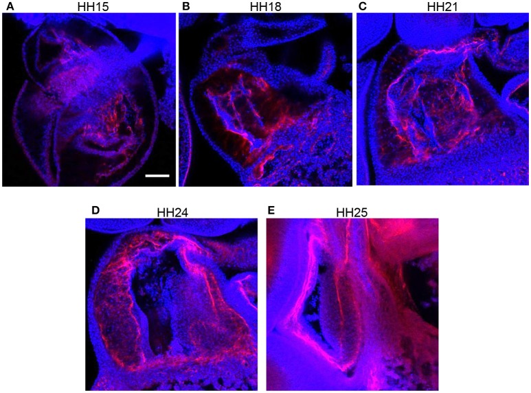
Normal blood flow is essential for proper heart formation during embryonic development, as abnormal hemodynamic load (blood pressure and shear stress) results in cardiac defects seen in congenital heart disease. However, the progressive detrimental remodeling processes that relate altered blood flow to cardiac defects remain unclear. Endothelial-mesenchymal cell transition is one of the many complex developmental events involved in transforming the early embryonic outflow tract into the aorta, pulmonary trunk, interventricular septum, and semilunar valves. This study elucidated the effects of increased hemodynamic load on endothelial-mesenchymal transition remodeling of the outflow tract cushions . Outflow tract banding was used to increase hemodynamic load in the chicken embryo heart between Hamburger and Hamilton stages 18 and 24. Increased hemodynamic load induced increased cell density in outflow tract cushions, fewer cells along the endocardial lining, endocardium junction disruption, and altered periostin expression as measured by confocal microscopy analysis. In addition, 3D focused ion beam scanning electron microscopy analysis determined that a portion of endocardial cells adopted a migratory shape after outflow tract banding that is more irregular, elongated, and with extensive cellular projections compared to normal cells. Proteomic mass-spectrometry analysis quantified altered protein composition after banding that is consistent with a more active stage of endothelial-mesenchymal transition. Outflow tract banding enhances the endothelial-mesenchymal transition phenotype during formation of the outflow tract cushions, suggesting that endothelial-mesenchymal transition is a critical developmental process that when disturbed by altered blood flow gives rise to cardiac malformation and defects.
正常血流对于胚胎发育过程中心脏的正常形成至关重要,因为异常的血流动力学负荷(血压和剪切应力)会导致先天性心脏病中出现的心脏缺陷。然而,将血流改变与心脏缺陷相关联的渐进性有害重塑过程仍不清楚。内皮-间充质细胞转变是将早期胚胎流出道转变为主动脉、肺动脉干、室间隔和半月瓣所涉及的众多复杂发育事件之一。本研究阐明了血流动力学负荷增加对流出道垫内皮-间充质转变重塑的影响。在汉堡和汉密尔顿18至24期之间,采用流出道结扎术增加鸡胚心脏的血流动力学负荷。通过共聚焦显微镜分析测量,增加的血流动力学负荷导致流出道垫中的细胞密度增加、内膜衬里的细胞减少、内膜连接破坏以及骨膜蛋白表达改变。此外,三维聚焦离子束扫描电子显微镜分析确定,与正常细胞相比,一部分内膜细胞在流出道结扎后呈现出更不规则、更长且具有广泛细胞突起的迁移形状。蛋白质组质谱分析定量了结扎后蛋白质组成的改变,这与内皮-间充质转变的更活跃阶段一致。流出道结扎增强了流出道垫形成过程中的内皮-间充质转变表型,表明内皮-间充质转变是一个关键的发育过程,当受到血流改变干扰时会导致心脏畸形和缺陷。