• 文献检索
  • 文档翻译
  • 深度研究
  • 学术资讯
  • Suppr Zotero 插件Zotero 插件
  • 邀请有礼
  • 套餐&价格
  • 历史记录
应用&插件
Suppr Zotero 插件Zotero 插件浏览器插件Mac 客户端Windows 客户端微信小程序
定价
高级版会员购买积分包购买API积分包
服务
文献检索文档翻译深度研究API 文档MCP 服务
关于我们
关于 Suppr公司介绍联系我们用户协议隐私条款
关注我们

Suppr 超能文献

核心技术专利:CN118964589B侵权必究
粤ICP备2023148730 号-1Suppr @ 2026

文献检索

告别复杂PubMed语法,用中文像聊天一样搜索,搜遍4000万医学文献。AI智能推荐,让科研检索更轻松。

立即免费搜索

文件翻译

保留排版,准确专业,支持PDF/Word/PPT等文件格式,支持 12+语言互译。

免费翻译文档

深度研究

AI帮你快速写综述,25分钟生成高质量综述,智能提取关键信息,辅助科研写作。

立即免费体验

乳腺癌体内分子成像的生物标志物:治疗前的F-FDG PET可预测患者预后,治疗前的DWI-MR可预测对新辅助化疗的反应。

Biomarkers from in vivo molecular imaging of breast cancer: pretreatment F-FDG PET predicts patient prognosis, and pretreatment DWI-MR predicts response to neoadjuvant chemotherapy.

作者信息

Gallivanone Francesca, Panzeri Marta Maria, Canevari Carla, Losio Claudio, Gianolli Luigi, De Cobelli Francesco, Castiglioni Isabella

机构信息

Institute of Molecular Bioimaging and Physiology, National Research Council (IBFM-CNR), Via Fratelli Cervi 93, Segrate, 20090, Milan, Italy.

Department of Radiology, Centre for Experimental Imaging, San Raffaele Scientific Institute, Milan, Italy.

出版信息

MAGMA. 2017 Aug;30(4):359-373. doi: 10.1007/s10334-017-0610-7. Epub 2017 Feb 28.

DOI:10.1007/s10334-017-0610-7
PMID:28246950
原文链接:https://pmc.ncbi.nlm.nih.gov/articles/PMC5524876/
Abstract

OBJECTIVE

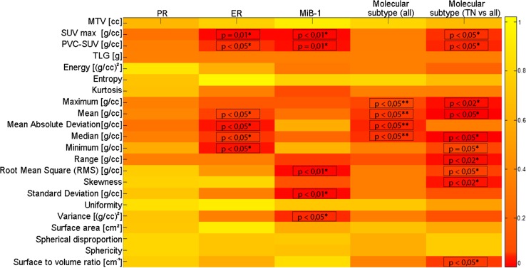
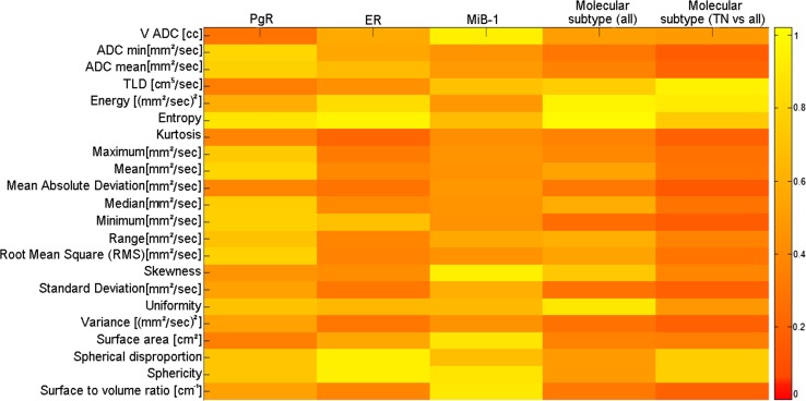
Human cancers display intra-tumor phenotypic heterogeneity and recent research has focused on developing image processing methods extracting imaging descriptors to characterize this heterogeneity. This work assesses the role of pretreatment F-FDG PET and DWI-MR with respect to the prognosis and prediction of neoadjuvant chemotherapy (NAC) outcomes when image features are used to characterize primitive lesions from breast cancer (BC).

MATERIALS AND METHODS

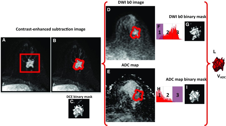
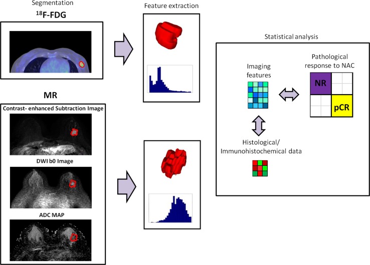
A retrospective protocol included 38 adult women with biopsy-proven BC. Patients underwent a pre-therapy F-FDG PET/CT whole-body study and a pre-therapy breast multi-parametric MR study. Patients were then referred for NAC treatment and then for surgical resection, with an evaluation of the therapy response. Segmentation methods were developed in order to identify functional volumes both on F-FDG PET images and ADC maps. Macroscopic and histogram features were extracted from the defined functional volumes.

RESULTS

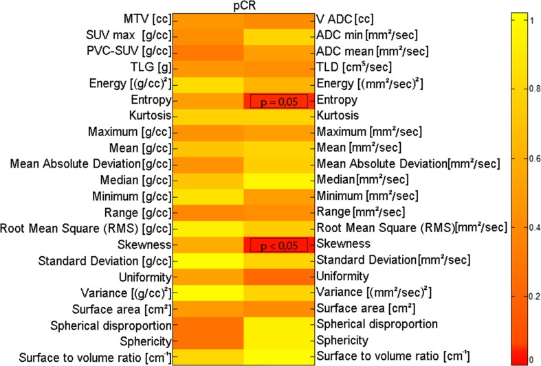
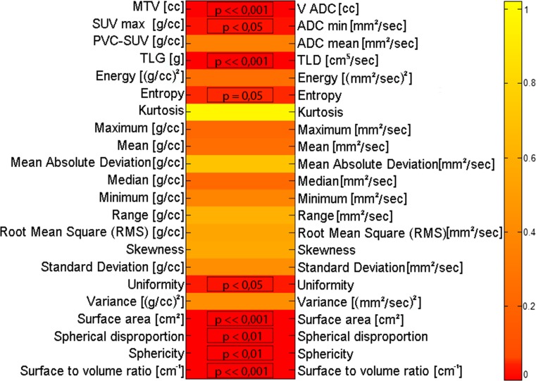
Our work demonstrates that macroscopic and histogram features from F-FDG PET are able to biologically characterize primitive BC, and define the prognosis. In addition, histogram features from ADC maps are able to predict the response to NAC.

CONCLUSION

Our work suggests that pre-treatment F-FDG PET and pre-treatment DWI-MR provide useful complementary information for biological characterization and NAC response prediction in BC.

摘要

目的

人类癌症表现出肿瘤内表型异质性,近期研究聚焦于开发图像处理方法来提取成像描述符以表征这种异质性。当使用图像特征来表征乳腺癌(BC)的原始病变时,本研究评估了治疗前F-FDG PET和DWI-MR在新辅助化疗(NAC)结果的预后和预测方面的作用。

材料与方法

一项回顾性研究纳入了38例经活检证实为BC的成年女性。患者在治疗前进行了F-FDG PET/CT全身检查和乳腺多参数MR检查。然后患者接受NAC治疗,随后进行手术切除,并对治疗反应进行评估。开发了分割方法以在F-FDG PET图像和ADC图上识别功能体积。从定义的功能体积中提取宏观和直方图特征。

结果

我们的研究表明,F-FDG PET的宏观和直方图特征能够从生物学上表征原始BC并确定预后。此外,ADC图的直方图特征能够预测对NAC的反应。

结论

我们的研究表明,治疗前F-FDG PET和治疗前DWI-MR为BC的生物学表征和NAC反应预测提供了有用的补充信息。

https://cdn.ncbi.nlm.nih.gov/pmc/blobs/2210/5524876/c11cde7cf599/10334_2017_610_Fig6_HTML.jpg
https://cdn.ncbi.nlm.nih.gov/pmc/blobs/2210/5524876/0b117f0b6fd9/10334_2017_610_Fig1_HTML.jpg
https://cdn.ncbi.nlm.nih.gov/pmc/blobs/2210/5524876/ee5d2102286e/10334_2017_610_Fig2_HTML.jpg
https://cdn.ncbi.nlm.nih.gov/pmc/blobs/2210/5524876/c6eef5238b61/10334_2017_610_Fig3_HTML.jpg
https://cdn.ncbi.nlm.nih.gov/pmc/blobs/2210/5524876/4f62d26d4316/10334_2017_610_Fig4_HTML.jpg
https://cdn.ncbi.nlm.nih.gov/pmc/blobs/2210/5524876/b1193deba5e1/10334_2017_610_Fig5_HTML.jpg
https://cdn.ncbi.nlm.nih.gov/pmc/blobs/2210/5524876/c11cde7cf599/10334_2017_610_Fig6_HTML.jpg

相似文献

1
Biomarkers from in vivo molecular imaging of breast cancer: pretreatment F-FDG PET predicts patient prognosis, and pretreatment DWI-MR predicts response to neoadjuvant chemotherapy.乳腺癌体内分子成像的生物标志物:治疗前的F-FDG PET可预测患者预后,治疗前的DWI-MR可预测对新辅助化疗的反应。
MAGMA. 2017 Aug;30(4):359-373. doi: 10.1007/s10334-017-0610-7. Epub 2017 Feb 28.
2
Baseline [F]FDG PET/CT and MRI first-order breast tumor features do not improve pathological complete response prediction to neoadjuvant chemotherapy.基线 [F]FDG PET/CT 和 MRI 一级乳腺癌肿瘤特征不能改善新辅助化疗的病理完全缓解预测。
Eur J Nucl Med Mol Imaging. 2024 Oct;51(12):3709-3718. doi: 10.1007/s00259-024-06815-6. Epub 2024 Jun 26.
3
Predicting neo-adjuvant chemotherapy response and progression-free survival of locally advanced breast cancer using textural features of intratumoral heterogeneity on F-18 FDG PET/CT and diffusion-weighted MR imaging.利用 F-18 FDG PET/CT 和弥散加权磁共振成像肿瘤内异质性的纹理特征预测局部晚期乳腺癌新辅助化疗反应和无进展生存期。
Breast J. 2019 May;25(3):373-380. doi: 10.1111/tbj.13032. Epub 2018 Mar 30.
4
Comparison of diffusion-weighted MR imaging and FDG PET/CT to predict pathological complete response to neoadjuvant chemotherapy in patients with breast cancer.比较扩散加权磁共振成像和 FDG PET/CT 预测乳腺癌患者新辅助化疗的病理完全缓解。
Eur Radiol. 2012 Jan;22(1):18-25. doi: 10.1007/s00330-011-2236-x. Epub 2011 Aug 16.
5
Baseline Tumor 18F-FDG Uptake and Modifications After 2 Cycles of Neoadjuvant Chemotherapy Are Prognostic of Outcome in ER+/HER2- Breast Cancer.基线肿瘤18F-FDG摄取及新辅助化疗2周期后的变化对雌激素受体阳性/人表皮生长因子受体2阴性乳腺癌预后具有预测价值。
J Nucl Med. 2015 Jun;56(6):824-31. doi: 10.2967/jnumed.115.154138. Epub 2015 Apr 16.
6
FDG-PET/CT and MRI for Evaluation of Pathologic Response to Neoadjuvant Chemotherapy in Patients With Breast Cancer: A Meta-Analysis of Diagnostic Accuracy Studies.氟代脱氧葡萄糖正电子发射断层扫描/计算机断层扫描(FDG-PET/CT)与磁共振成像(MRI)用于评估乳腺癌患者新辅助化疗后的病理反应:诊断准确性研究的荟萃分析
Oncologist. 2016 Aug;21(8):931-9. doi: 10.1634/theoncologist.2015-0353. Epub 2016 Jul 8.
7
Correlation of simultaneously acquired diffusion-weighted imaging and 2-deoxy-[18F] fluoro-2-D-glucose positron emission tomography of pulmonary lesions in a dedicated whole-body magnetic resonance/positron emission tomography system.在专用全身磁共振/正电子发射断层扫描系统中对肺部病变进行同时采集的弥散加权成像和 2-脱氧-[18F]氟-2-D-葡萄糖正电子发射断层扫描的相关性研究。
Invest Radiol. 2013 May;48(5):247-55. doi: 10.1097/RLI.0b013e31828d56a1.
8
Combination of 18F-FDG PET/CT and diffusion-weighted MR imaging as a predictor of histologic response to neoadjuvant chemotherapy: preliminary results in osteosarcoma.18F-FDG PET/CT 与弥散加权磁共振成像联合预测骨肉瘤新辅助化疗的组织学反应:初步结果。
J Nucl Med. 2013 Jul;54(7):1053-9. doi: 10.2967/jnumed.112.115964. Epub 2013 May 13.
9
The value of pre- and post-neoadjuvant chemotherapy F-18 FDG PET/CT scans in breast cancer: comparison with MRI.新辅助化疗前后F-18 FDG PET/CT扫描在乳腺癌中的价值:与MRI的比较
Acta Radiol. 2018 Jan;59(1):41-49. doi: 10.1177/0284185117705011. Epub 2017 Apr 21.
10
Clinical implications of non-breast cancer related findings on FDG-PET/CT scan prior to neoadjuvant chemotherapy in patients with breast cancer.乳腺癌患者新辅助化疗前 FDG-PET/CT 扫描非乳腺癌相关发现的临床意义。
Breast Cancer Res Treat. 2024 Aug;206(3):585-594. doi: 10.1007/s10549-024-07331-9. Epub 2024 Jun 12.

引用本文的文献

1
Does FDG PETCT have a predictive value for neoadjuvant chemotherapy response in nonmetastatic breast cancer?氟代脱氧葡萄糖正电子发射断层扫描计算机断层扫描(FDG PETCT)对非转移性乳腺癌新辅助化疗反应是否具有预测价值?
Ir J Med Sci. 2025 Feb;194(1):31-36. doi: 10.1007/s11845-024-03856-6. Epub 2024 Dec 18.
2
Baseline [F]FDG PET/CT and MRI first-order breast tumor features do not improve pathological complete response prediction to neoadjuvant chemotherapy.基线 [F]FDG PET/CT 和 MRI 一级乳腺癌肿瘤特征不能改善新辅助化疗的病理完全缓解预测。
Eur J Nucl Med Mol Imaging. 2024 Oct;51(12):3709-3718. doi: 10.1007/s00259-024-06815-6. Epub 2024 Jun 26.
3

本文引用的文献

1
Metabolic Tumor Burden Assessed by Dual Time Point [F]FDG PET/CT in Locally Advanced Breast Cancer: Relation with Tumor Biology.双时间点[F]FDG PET/CT评估局部晚期乳腺癌的代谢肿瘤负荷:与肿瘤生物学的关系
Mol Imaging Biol. 2017 Aug;19(4):636-644. doi: 10.1007/s11307-016-1034-x.
2
Research needs in breast cancer.乳腺癌研究需求。
Ann Oncol. 2017 Feb 1;28(2):208-217. doi: 10.1093/annonc/mdw571.
3
Early Change in FDG-PET Signal and Plasma Cell-Free DNA Level Predicts Erlotinib Response in EGFR Wild-Type NSCLC Patients.氟代脱氧葡萄糖正电子发射断层扫描(FDG-PET)信号和血浆游离DNA水平的早期变化可预测表皮生长因子受体(EGFR)野生型非小细胞肺癌(NSCLC)患者对厄洛替尼的反应。
Prediction of pathological response after neoadjuvant chemotherapy using baseline FDG PET heterogeneity features in breast cancer.
使用乳腺癌基线 FDG PET 异质性特征预测新辅助化疗后的病理反应。
Br J Radiol. 2023 Jun 1;96(1146):20220655. doi: 10.1259/bjr.20220655. Epub 2023 Mar 3.
4
Radiogenomics, Breast Cancer Diagnosis and Characterization: Current Status and Future Directions.放射基因组学、乳腺癌诊断与特征分析:现状与未来方向
Methods Protoc. 2022 Oct 3;5(5):78. doi: 10.3390/mps5050078.
5
Invasive ductal breast cancer molecular subtype prediction by MRI radiomic and clinical features based on machine learning.基于机器学习,通过MRI影像组学和临床特征预测浸润性导管乳腺癌分子亚型
Front Oncol. 2022 Sep 12;12:964605. doi: 10.3389/fonc.2022.964605. eCollection 2022.
6
Factors affecting the value of diffusion-weighted imaging for identifying breast cancer patients with pathological complete response on neoadjuvant systemic therapy: a systematic review.影响弥散加权成像在新辅助全身治疗中识别达到病理完全缓解的乳腺癌患者价值的因素:一项系统评价
Insights Imaging. 2021 Dec 18;12(1):187. doi: 10.1186/s13244-021-01123-1.
7
How to Discriminate Lung Cancer From Benign Pulmonary Nodules and Masses? Usefulness of Diffusion-Weighted Magnetic Resonance Imaging With Apparent Diffusion Coefficient and Inside/Wall Apparent Diffusion Coefficient Ratio.如何鉴别肺癌与良性肺结节及肿块?基于表观扩散系数和内部/壁表观扩散系数比值的扩散加权磁共振成像的效用
Clin Med Insights Oncol. 2021 Jul 7;15:11795549211014863. doi: 10.1177/11795549211014863. eCollection 2021.
8
Advanced Imaging Analysis in Prostate MRI: Building a Radiomic Signature to Predict Tumor Aggressiveness.前列腺MRI的高级影像分析:构建用于预测肿瘤侵袭性的影像组学特征
Diagnostics (Basel). 2021 Mar 26;11(4):594. doi: 10.3390/diagnostics11040594.
9
Relationships and Qualitative Evaluation Between Diffusion-Weighted Imaging and Pathologic Findings of Resected Lung Cancers.肺癌切除标本的扩散加权成像与病理结果之间的关系及定性评估
Cancers (Basel). 2020 May 8;12(5):1194. doi: 10.3390/cancers12051194.
10
AI-based applications in hybrid imaging: how to build smart and truly multi-parametric decision models for radiomics.基于人工智能的混合成像应用:如何为放射组学构建智能且真正多参数的决策模型。
Eur J Nucl Med Mol Imaging. 2019 Dec;46(13):2673-2699. doi: 10.1007/s00259-019-04414-4. Epub 2019 Jul 11.
Transl Oncol. 2016 Dec;9(6):505-511. doi: 10.1016/j.tranon.2016.09.003. Epub 2016 Oct 29.
4
18F-fludrodeoxyglucose maximal standardized uptake value and metabolic tumor burden are associated with major chemotherapy-related tumor markers in NSCLC patients.18F-氟脱氧葡萄糖最大标准化摄取值和代谢肿瘤负荷与非小细胞肺癌患者主要化疗相关肿瘤标志物相关。
Onco Targets Ther. 2016 Oct 14;9:6315-6324. doi: 10.2147/OTT.S113832. eCollection 2016.
5
Whole-Tumor Quantitative Apparent Diffusion Coefficient Histogram and Texture Analysis to Predict Gleason Score Upgrading in Intermediate-Risk 3 + 4 = 7 Prostate Cancer.全肿瘤定量表观扩散系数直方图和纹理分析预测中危3+4=7前列腺癌的Gleason评分升级
AJR Am J Roentgenol. 2016 Apr;206(4):775-82. doi: 10.2214/AJR.15.15462. Epub 2016 Feb 2.
6
Diagnostic performance of texture analysis on MRI in grading cerebral gliomas.MRI纹理分析在脑胶质瘤分级中的诊断性能
Eur J Radiol. 2016 Apr;85(4):824-9. doi: 10.1016/j.ejrad.2016.01.013. Epub 2016 Jan 21.
7
CT-based compartmental quantification of adipose tissue versus body metrics in colorectal cancer patients.基于CT的结直肠癌患者脂肪组织与身体指标的分区定量分析
Eur Radiol. 2016 Nov;26(11):4131-4140. doi: 10.1007/s00330-016-4231-8. Epub 2016 Feb 6.
8
Texture analysis on parametric maps derived from dynamic contrast-enhanced magnetic resonance imaging in head and neck cancer.头颈部癌动态对比增强磁共振成像衍生参数图的纹理分析
World J Radiol. 2016 Jan 28;8(1):90-7. doi: 10.4329/wjr.v8.i1.90.
9
FDG PET/CT texture analysis for predicting the outcome of lung cancer treated by stereotactic body radiation therapy.用于预测立体定向体部放射治疗的肺癌患者预后的氟代脱氧葡萄糖正电子发射断层显像/计算机断层扫描纹理分析
Eur J Nucl Med Mol Imaging. 2016 Jul;43(8):1453-60. doi: 10.1007/s00259-016-3314-8. Epub 2016 Jan 30.
10
Diffusion-weighted imaging and (18) F-fluorodeoxyglucose positron emission tomography/computed tomography in breast cancer: Correlation of the apparent diffusion coefficient and maximum standardized uptake values with prognostic factors.扩散加权成像及(18)F-氟脱氧葡萄糖正电子发射断层扫描/计算机断层扫描在乳腺癌中的应用:表观扩散系数和最大标准化摄取值与预后因素的相关性
J Magn Reson Imaging. 2016 Jun;43(6):1434-44. doi: 10.1002/jmri.25112. Epub 2015 Dec 10.