Nau Christoph, Henrich Dirk, Seebach Caroline, Schröder Katrin, Barker John H, Marzi Ingo, Frank Johannes
Department of Trauma, Hand and Reconstructive Surgery, Johann Wolfgang Goethe‑University, Frankfurt/Main, Germany.
Institute for Cardiovascular Physiology, Goethe-University, Frankfurt/Main, Germany.
Int J Mol Med. 2017 Apr;39(4):907-917. doi: 10.3892/ijmm.2017.2901. Epub 2017 Feb 21.
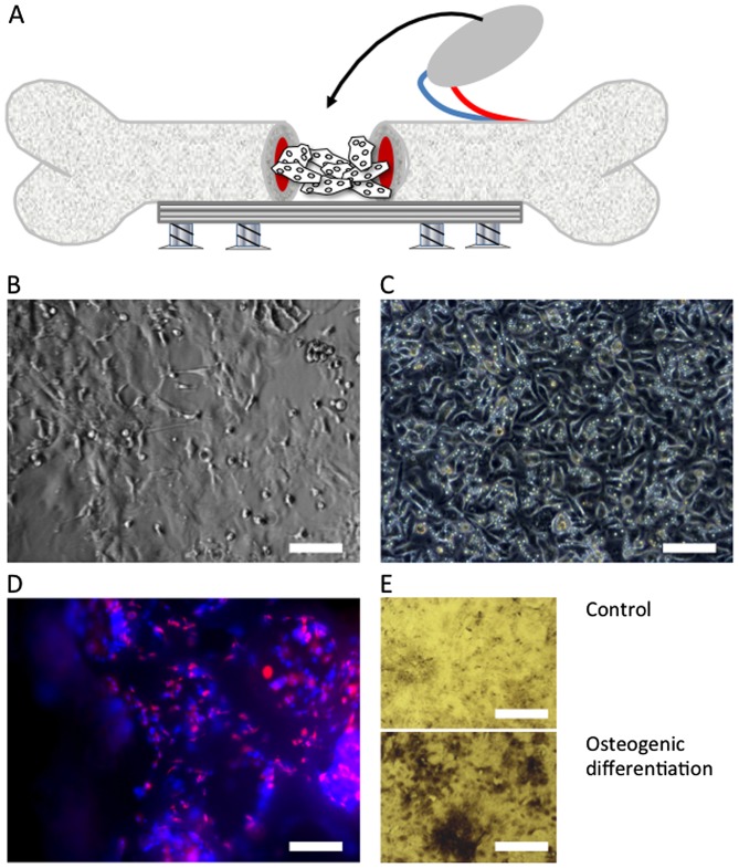
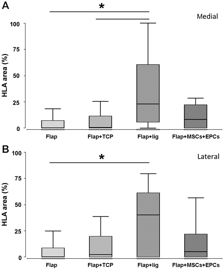
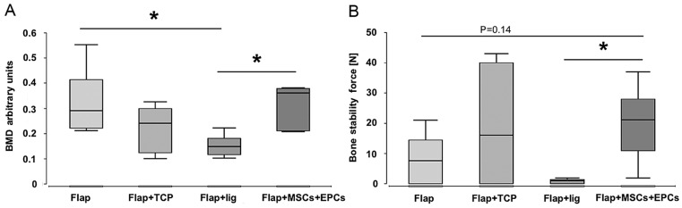
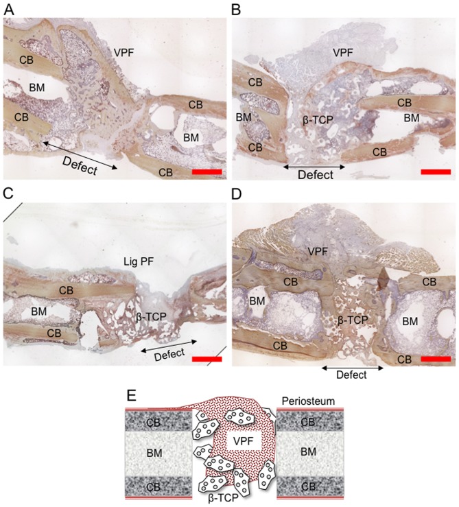
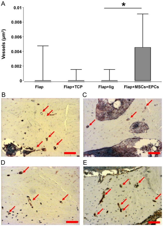
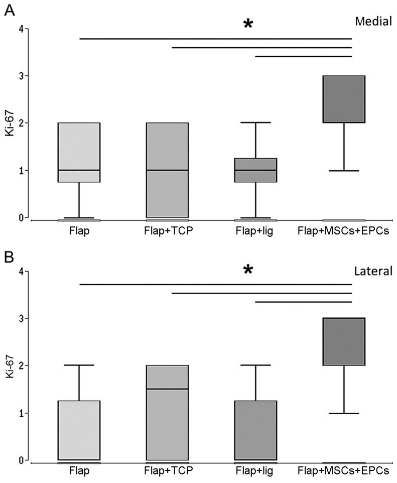
Vascularized periosteal flaps are used for complex cases if the reconstruction of large bone defects is necessary in modern trauma and orthopedic surgery. In this study, we combined this surgical procedure with β‑TCP scaffold and mesenchymal stem cells (MSCs) + endothelial progenitor cells (EPCs) as a tissue engineering approach to obtain optimum conditions for bone healing in rats. A critical size femoral defect was created in 80 rats allocated into 4 groups. Defects were treated according to the following protocol: i) vascularized periosteal flap alone; ⅱ) vascularized periosteal flap + β‑TCP scaffold; ⅲ) vascularized periosteal flap + β‑TCP scaffold + ligated vascular pedicle; and ⅳ) vascularized periosteal flap + β‑TCP scaffold + MSCs/EPCs. After 8 weeks, femur bones were extracted and analyzed for new bone formation, vascularization, proliferation and inflammatory processes and strength. Bone mineral density (BMD) and biomechanical stability at week 8 were highest in group 4 (flap + β‑TCP scaffold + MSCs/EPCs) compared to all the other groups. Stability was significantly higher in group 4 (flap + β‑TCP scaffold + MSCs/EPCs) in comparison to group 3 (ligated flap + β‑TCP scaffold). BMD was found to be significantly lower in group 3 (ligated flap + β‑TCP scaffold) compared to group 1 (flap) and group 4 (flap + β‑TCP scaffold + MSCs/EPCs). The highest density of blood vessels was observed in group 4 (flap + β‑TCP + MSCs/EPCs) and the values were significantly increased in comparison to group 3 (ligated flap), but not to group 1 (flap) and group 2 (flap + β‑TCP). The highest amounts of proliferating cells were observed in group 4 (flap + β‑TCP scaffold + MSC/EPCs). The percentage of proliferating cells was significantly higher in group 4 (flap + β‑TCP scaffold + MSCs/EPCs) in comparison to all the other groups after 8 weeks. Our data thus indicate that critical size defect healing could be improved if MSCs/EPCs are added to β-TCP scaffold in combination with a periosteal flap. Even after 8 weeks, the amount of proliferating cells was increased. The flap blood supply is essential for bone healing and the reduction of inflammatory processes.
在现代创伤和骨科手术中,如果需要重建大的骨缺损,血管化骨膜瓣可用于复杂病例。在本研究中,我们将这种手术方法与β - 磷酸三钙(β - TCP)支架以及间充质干细胞(MSCs)+内皮祖细胞(EPCs)相结合,作为一种组织工程方法,以获得大鼠骨愈合的最佳条件。在80只大鼠中制造了临界尺寸的股骨缺损,并将其分为4组。根据以下方案治疗缺损:i)单独使用血管化骨膜瓣;ⅱ)血管化骨膜瓣 + β - TCP支架;ⅲ)血管化骨膜瓣 + β - TCP支架 + 结扎血管蒂;ⅳ)血管化骨膜瓣 + β - TCP支架 + MSCs/EPCs。8周后,取出股骨并分析新骨形成、血管化、增殖和炎症过程以及强度。与所有其他组相比,第4组(骨膜瓣 + β - TCP支架 + MSCs/EPCs)在第8周时的骨密度(BMD)和生物力学稳定性最高。与第3组(结扎骨膜瓣 + β - TCP支架)相比,第4组(骨膜瓣 + β - TCP支架 + MSCs/EPCs)的稳定性显著更高。发现第3组(结扎骨膜瓣 + β - TCP支架)的BMD显著低于第1组(骨膜瓣)和第4组(骨膜瓣 + β - TCP支架 + MSCs/EPCs)。在第4组(骨膜瓣 + β - TCP + MSCs/EPCs)中观察到最高的血管密度,与第3组(结扎骨膜瓣)相比,该值显著增加,但与第1组(骨膜瓣)和第2组(骨膜瓣 + β - TCP)相比则没有增加。在第4组(骨膜瓣 + β - TCP支架 + MSC/EPCs)中观察到增殖细胞数量最多。8周后,与所有其他组相比,第4组(骨膜瓣 + β - TCP支架 + MSCs/EPCs)中增殖细胞的百分比显著更高。因此,我们的数据表明,如果将MSCs/EPCs添加到β - TCP支架并结合骨膜瓣,临界尺寸缺损的愈合可以得到改善。即使在8周后,增殖细胞的数量也有所增加。骨膜瓣的血液供应对于骨愈合和减少炎症过程至关重要。